We evaluate the aortic valve for pathology on every patient we scan. It’s easy to forget the basic concept of anatomy and the functions each part of the valve plays in opening & closing of the leaflets. This blog is going to be a refresher and cover the basic anatomy of the aortic valve!
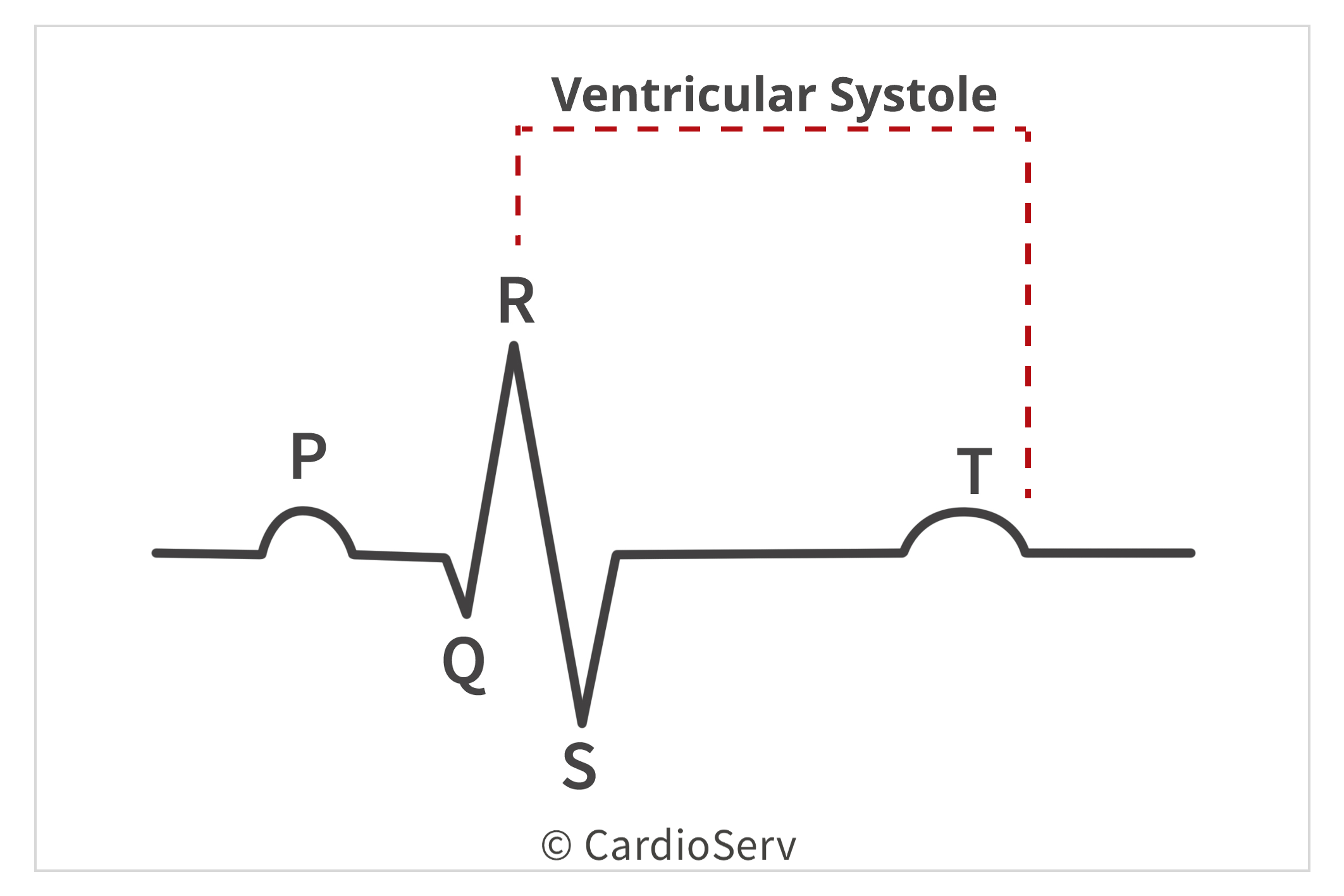
The aortic valve’s main function is to act as a gateway for blood to exit out of the ventricle during systole and push through the aorta for the body to receive oxygenated blood. Ventricular systole is a very short amount of time during the cardiac cycle for the leaflets to open and close, allowing blood to exit out of the heart.
The aortic valve is composed of 4 key parts:
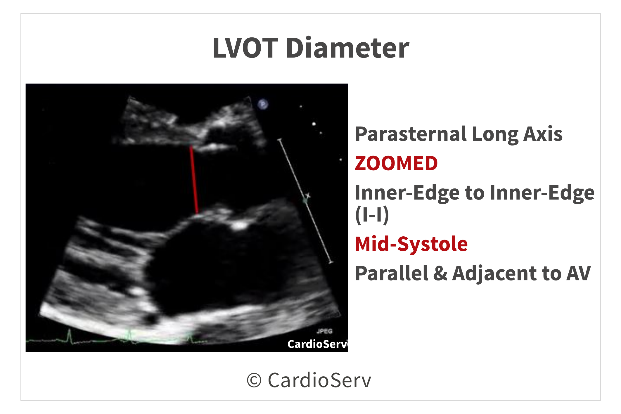
The aortic annulus is a functioning ring-like structure that anchors the base hinge-points of the aortic leaflets. We measure the aortic annulus diameter between the hinge points of the valve leaflets, commonly referred to as the LVOT diameter.
The aortic root is composed of 3 parts:
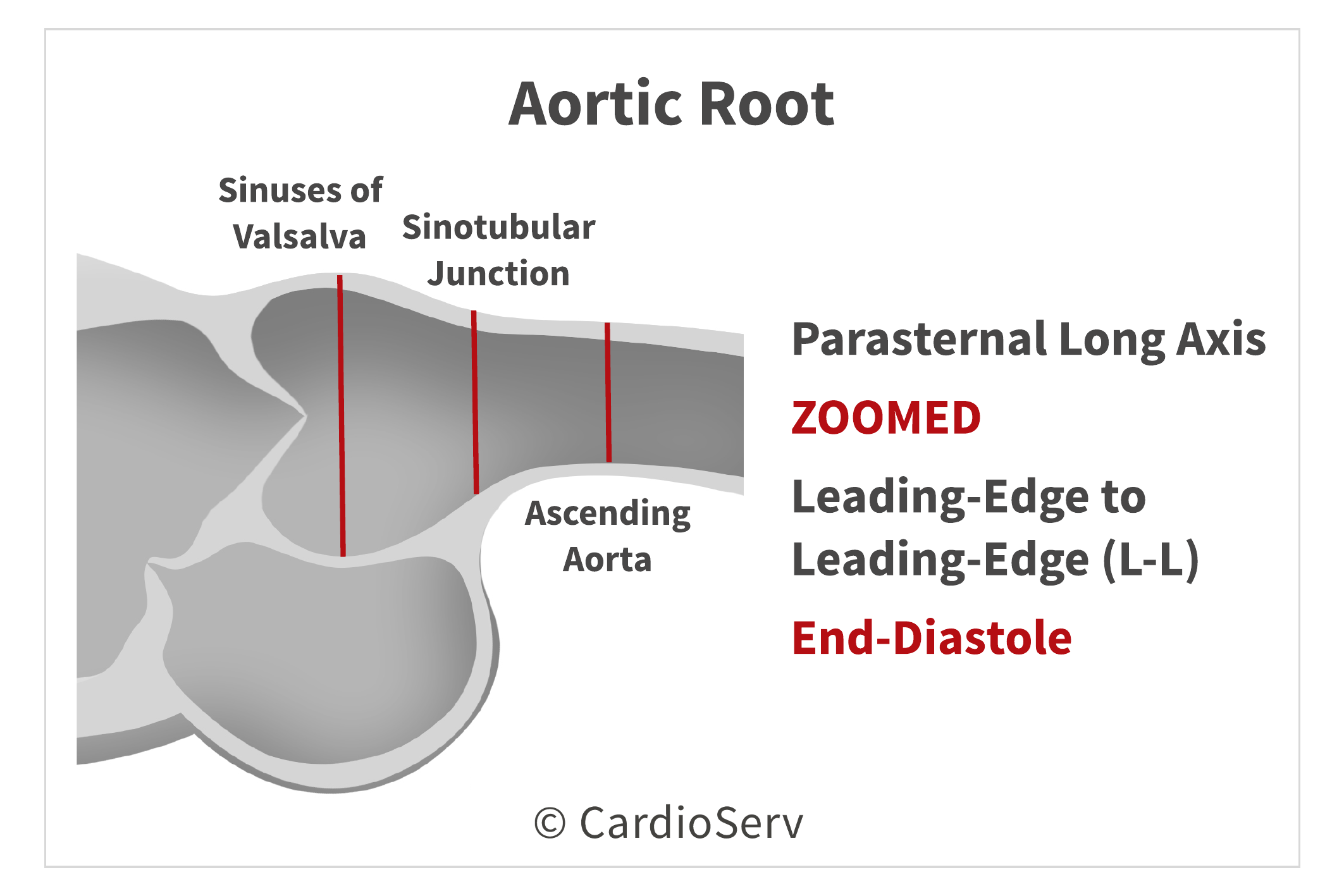
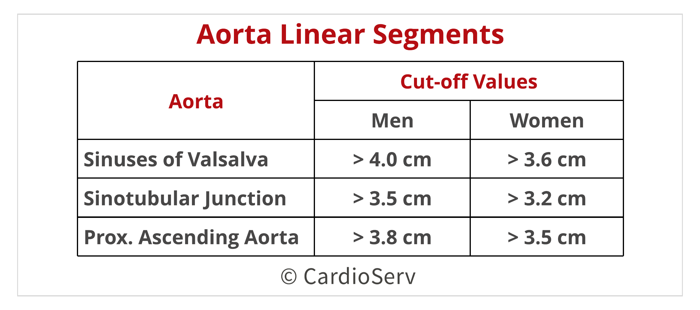
When measuring various parts of the aortic root, it is vital as a department to decide on 1 specific location to measure and label accordingly to specific site. Reference values are specific to each segment within the aortic root.
The ASE explains 2D measurements are preferred over M-Mode calculations due to cardiac movement that can easily alter the location of the cursor. 2D linear measurement ensures the maximum diameter is being obtained.
The sinuses of Valsalva are outward pouches within the aortic root where the coronary arteries arise from. They act as a reservoir of blood to supplement the coronaries during diastole. During systole, the outward pouches act as support for the leaflet cusps.
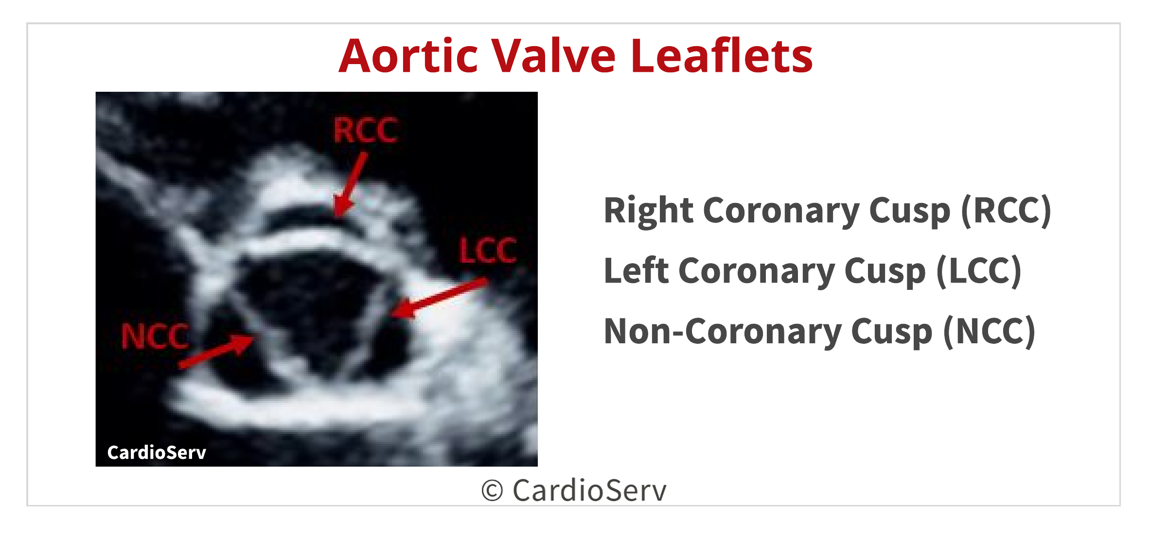
The aortic valve is composed of 3 leaflets. Each leaflet is named according to the sinuses of Valsalva outward pouch it overlies. Since the outward pouches are associated with the coronary artery, the leaflet cusps are named accordingly:
Therefore, we can associate the RCC with the right coronary artery and the LCC with the left coronary artery. Each cusp has two free edges, which is shared with the neighboring cusp.
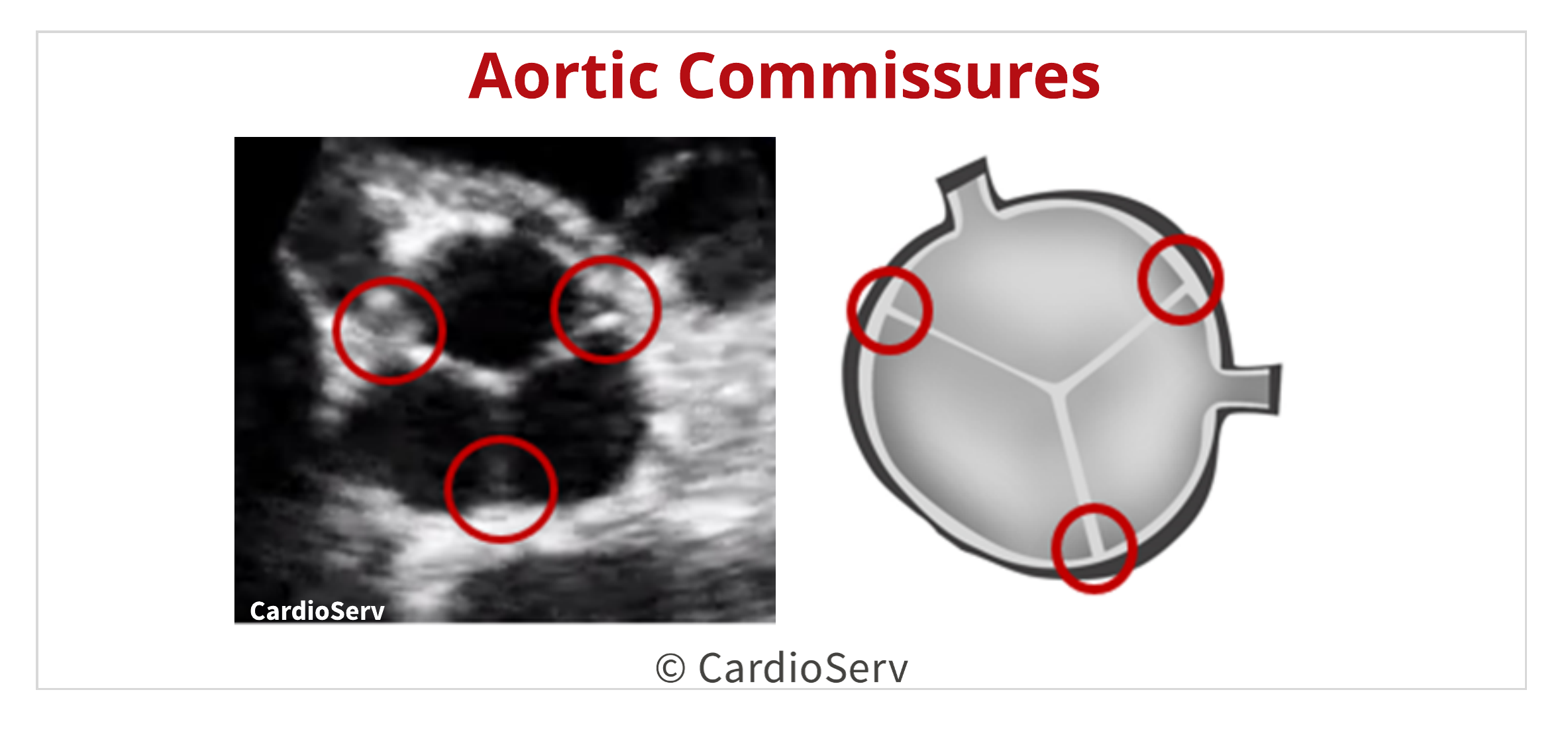
The aortic valve is composed of 3 commissures. A commissure is the space or area between each anchored leaflet to the aortic wall. They act as support to the base structure of the cusps.
Knowing the basic anatomy of the aortic valve allows us to better evaluate and diagnose patients with pathology. This blog provides the necessary steps to understanding, evaluating and measuring the structure that make up the aortic valve.


Andrea Fields MHA, RDCS
Stay Connected: LinkedIn, Facebook, Twitter, Instagram
References:
Lang, R. M., MD, Badano, L. P., MD, & Mor-Avi, V., PhD. (2015). Recommendations for Cardiac Chamber Quantification by Echocardiography in Adults: An Update from the American Society of Echocardiography and the European Association of Cardiovascular Imaging. JASE, 28(1), 1-53. Retrieved March 1, 2017, from http://asecho.org/wordpress/wp-content/uploads/2015/01/ChamberQuantification2015.pdf


Feb
2018
Feb
2018
Feb
2018
Mar
2018
Jun
2018
Nov
2018
Jan
2019
Mar
2020