fbpx
Written by Andrea Fields MHA, RDCS

3 Doppler Techniques for Evaluation of MR!

Last week we discussed 3 measures to use with color Doppler for evaluating the presence of mitral regurgitation (MR). If you missed last week’s blog, or would like to refresh, you can find them here:

  1. Mitral Valve Anatomy: Name 5 Components! 
  2. Finally… Mitral Valve Orientation Explained!
  3. Smart Strategies for Determining MR Mechanisms
  4. Ultimate Guide to Acute vs. Chronic MR
  5. Essential Steps to Evaluating MR Etiologies
  6. Secondary MR: Evaluating Mitral Valve Tethering
  7. FYI: Temporal Variation & Blood Pressure Matters for MR!
  8. 7 Factors that Influence Color Doppler Appearance 
  9. 3 Essential Color Doppler Measurements for MR!

This week, we are going to cover 3 qualitative techniques with the presence of MR using pulsed-waved (PW) Doppler and continuous-waved (CW) Doppler! By the end of this blog, we hope to have a better understanding of how these qualitative methods help us in determining the severity of MR:

  1. CW Doppler MR Jet Velocity
  2. PW Doppler Mitral Valve Inflow
  3. PW Doppler of Pulmonary Veins

CW Doppler Jet

Mitral Regurgitation CW Doppler

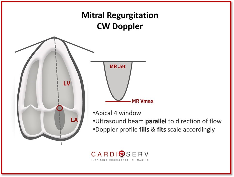
The profile of the Doppler jet can tell us a lot of information regarding hemodynamics and transvalvular flow rates. Even if MR is not detected with color Doppler, it is still essential to prove this with placing a CW Doppler through the mitral valve (MV) to demonstrate absence of flow below the baseline (TTE).

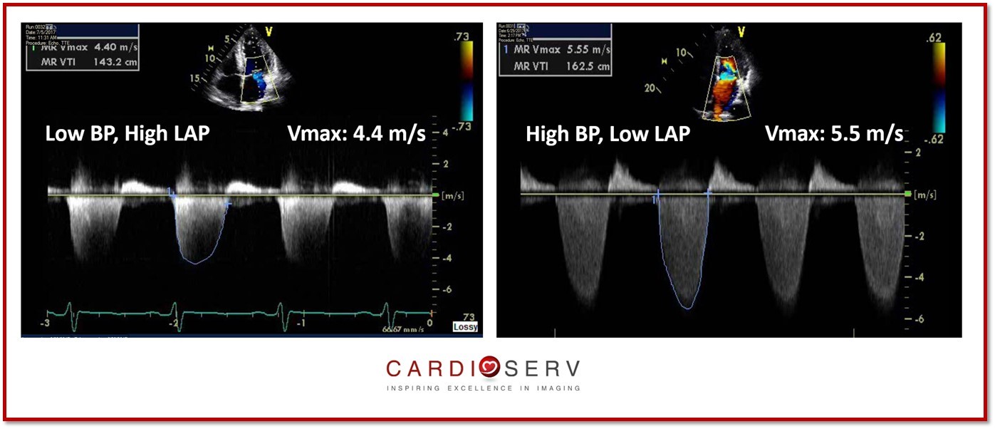
The max velocity of the MR jet does not provide us information on the severity of the MR. It does provide us with information regarding the hemodynamics of the LV & LA in regards to the MR. The max velocity of the jet represents the instantaneous systolic pressure gradient between the LV and LA.

It is common for the MR jet velocity to range between 4-6 m/s. This can allow us to easily analyze different scenarios:

  • MR Vmax 4 m/s : indicate a low BP and high LAP
  • MR Vmax 6 m/s : indicate a high BP and low LAP

Mitral Regurgitation CW Jet Velocity

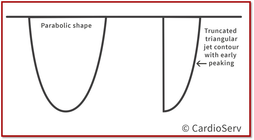
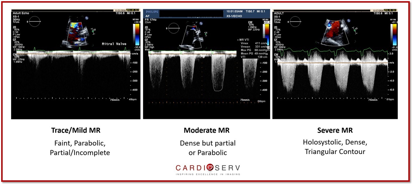
The contour and density of the jet can also provide qualitative information.

  • Contour: aids in identifying the severity of MR
  • Density: represents the number of RBC’s in the signal (amplitude & intensity)

Mitral Regurgitation Jet Profile

Mitral Regurgitation Jet Profile

PW Doppler MV Inflow

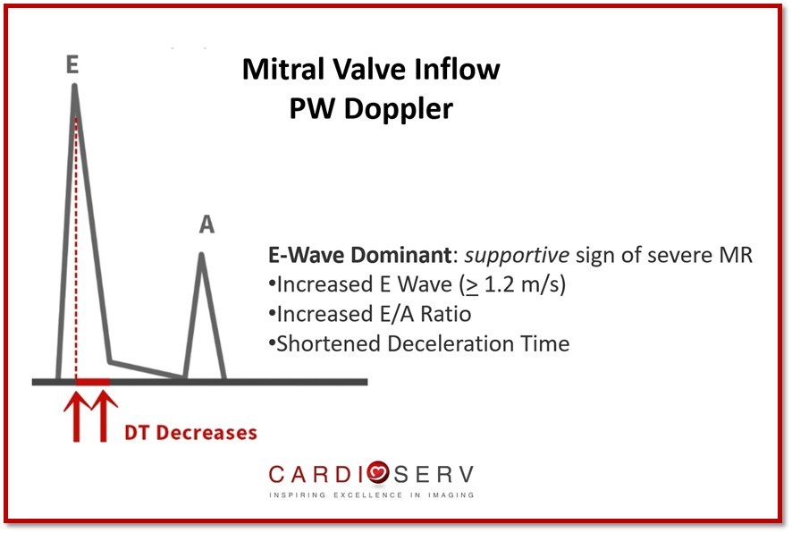
The inflow pattern and velocity can help us identify supportive signs of severe MR!

  • Apical 4 window
  • Ultrasound beam parallel to direction of flow
  • Doppler profile fills & fits scale accordingly
  • Gains adjusted for optional imaging

This velocity informs us of the forward stroke volume (SV) across the mitral valve. It can be increased if regurgitation is present.

  • E-Wave Dominantsupportive sign of severe MR
    • Increased E Wave (> 1.2 m/s)
    • Increased E/A Ratio
    • Shortened Decel Time
  • Reliable to assess primary MR
    • Challenge for secondary
    • Unable to differentiate between LV filling pressures or regurgitation
  • Dominant A-Wave inflow pattern excludes severe MR

Mitral Valve PW Inflow

PW Doppler Pulmonary Veins

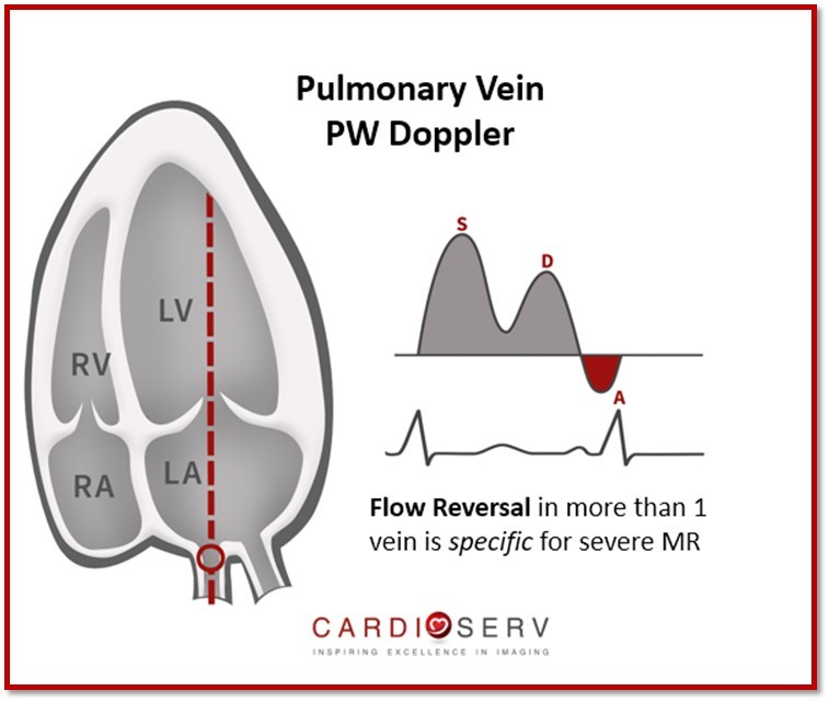
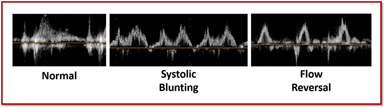
We can Doppler the pulmonary veins to evaluate for flow reversal and the hemodynamics due to MR.

  • Apical 4 window
  • Sample volume of Doppler 1 cm into pulmonary vein
  • Do not Doppler a vein that regurgitation is dumping into; attempt more than one vein in this case

Normal Pulmonary Vein Waveform

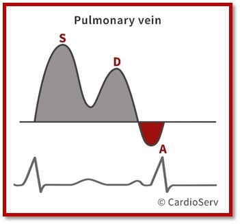
Components of pulmonary vein waveform:

  1. S Wave: systolic
  2. D Wave: diastolic
  3. A Wave: peak reversal flow
  4. Ar Duration: time of peak reversal flow (atrial contraction)

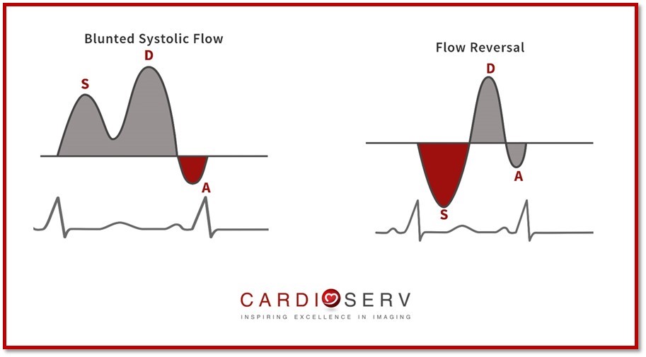
Flow reversal pattern is present, there is > 85% probability of severe MR.

Pulmonary Veins for MR

Items to note:

  • Systolic blunted flow does not automatically indicate moderate MR- could be the result of diastolic dysfunction.
  • Normal flow pattern suggest low LAP & non-severe MR
  • Less valuable in secondary MR due to unable to exclude diastolic dysfunction

Pulmonary Vein Waveforms

Pulmonary Vein Flow

Summary

We hope that you can utilize these qualitative and semi-quantitative measures within your evaluation of severity of MR! In this blog, we covered information in regards to MR about:

  • CW Doppler of MR Jet
  • PW Doppler of Mitral Valve Inflow
  • PW Doppler of Pulmonary Veins

Next week, we will start discussing quantitative methods to determining the severity of mitral regurgitation! We love to hear feedback and comments from our readers!

Andrea Fields MHA, RDCS

Stay Connected: LinkedInFacebookTwitterInstagram


References: 

Zoghbi, W. A., MD, FASE, & Adams, D., RCS, RDCS, FASE. (2017). Recommendations for Noninvasive Evaluation of Native Valvular Regurgitation. JASE, 30, 4th ser., 1-69. Retrieved June 12, 2017.

ASE guidelinesCW DopplerMitral RegurgitationMitral ValveMitral Valve InflowMR Jet VelocityPulmonary VeinsPW DopplerRegurgitation Quantification

Our Services

LET US KNOW WHAT YOU THINK...

Ayed
Thanks
Kathirvel
Well done, thanks
OSAMA ELKADI
grades of pulm reverse flow in relation of mr thanks
Tadesse Gemechuhow d
thank you
Samir srour
thanks
bassim
Great ....and I would like to say thanks for this nice work and informative subject
Alexander Hernandez
Useful tips
Teodosi
Great!Thanks!
Dr.Nazrul Islam
It's an excellent way of teaching.
Rosemary Beles Niesl
more infomation on tissue doppler
HAGUMAKUBANA OLIVIER
it is a very summarized but very effective. Thanks.
Carol
I have been on Hiatus for the last 2 years from my 16 yr CV Tech Career. I am now ready to jump back in and recertify. Thank you for all the great information and tips.
LAP Perera
Thankyou
Omar
Thank you very much Really useful
Saine
Thank you very much.It helps me to get more knowledge about MR and Pulmonary Vein Measurements.Million thanks to you all.

Your email will not be displayed.

Required *

Blog
Sign Up

Accreditation
services

Accreditation Help
Online Echo CMEs
Echo Accreditation Software
Free
Consultation

Search
for

Blog
Categories

Blog
Archive

Tags
Used

Show tags
Need Help? Share