Written by Andrea Fields MHA, RDCS
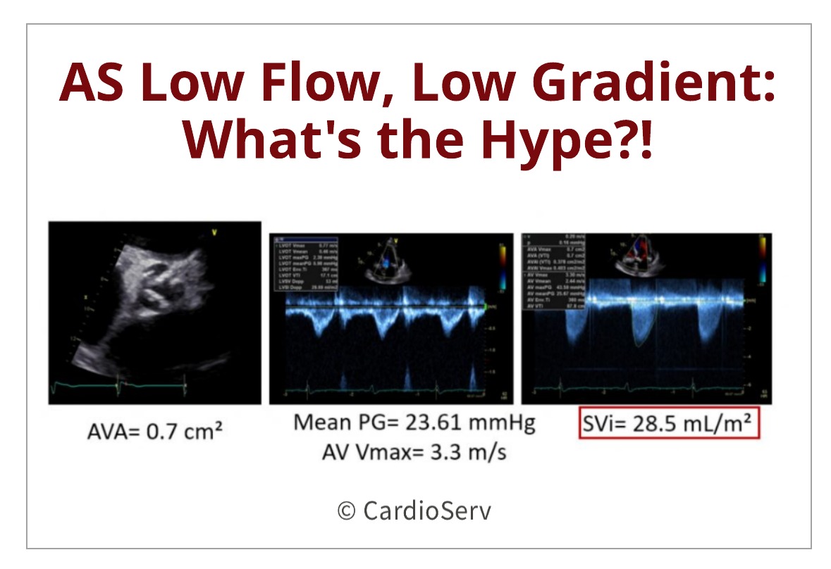
We've all been in this situation.... our patient appears to have a very tight, calcified aortic valve which visually appears to be moderate to severe aortic stenosis. As we scan through our protocol, we obtain a peak aortic valve velocity of 3.3 m/s and mean pressure gradient (PG) of 23.6 mmHg. The...
Written by Andrea Fields MHA, RDCS
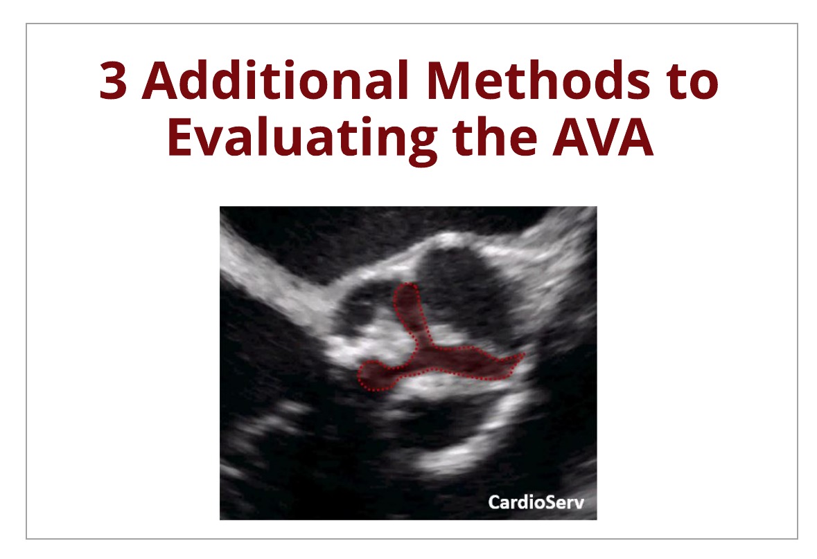
Last week we broke down the continuity equation to evaluate the severity of aortic stenosis. This week, we are going to discuss other methods that can be used to evaluate the severity of aortic stenosis. This blog will cover the following methods:
AVA Planimetry
Indexed Continuity...
Written by Andrea Fields MHA, RDCS
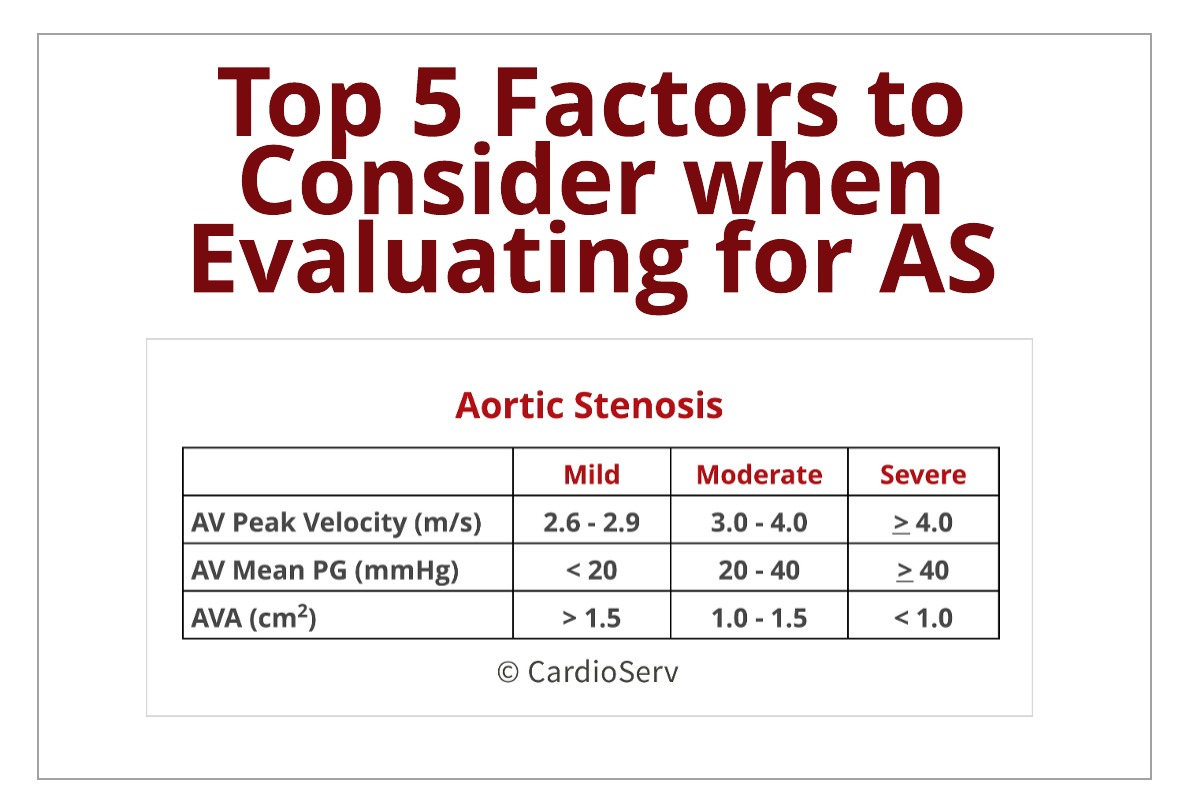
Last week we broke down the methodology of the continuity equation to calculate the aortic valve area (AVA). If you missed it, you can find it HERE. The ASE guideline recommends using the continuity equation calculated AVA, peak aortic jet velocity and mean pressure gradient to evaluate and...
Written by Andrea Fields MHA, RDCS
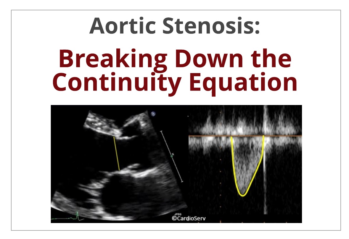
The evaluation and screening of aortic stenosis is a routine calculation performed on all complete echocardiograms. The detailed evaluation involves multiple key parameters that make up the equation that determines the aortic valve area (AVA). With echo being the gold-standard to evaluating and...
Written by Judith Buckland, MBA, RDCS, FASE
If you work in a cardiology lab you know the challenges of scheduling patients for both their echocardiogram and nuclear stress test. The stress test is already a long day for the patient and so often, in an effort to accommodate our patients, we schedule both the echo and the stress test on the...
 Cardioserv Blog