Written by Judith Buckland, RDCS, FASE, MBA
How could I improve this image?
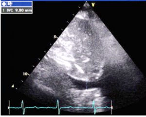
Modality: Transthoracic Echocardiography
View: Subcostal long axis IVC
Answer:
The image could be optimized to elongate the IVC to ensure the correct diameter is measured.
Tips for Measuring the IVC:
- Long axis view of the IVC
- Optimize image so that the IVC is open uniformly both proximally and distally where it can be seen emptying into the right atrium
- Slight angulation or sweeping through the IVC helps find the true midline diameter
- The diameter of the IVC should be measured 1-2 cm from the right atrium
- The IVC should be measured Inner-to-Inner Edge (I-I)
- Be sure to measure perpendicular to the vessel
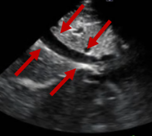
Correct Technique:
Notice how the IVC is fully opened up to correctly display a uniformed opening both proximally and distally. For more information on correct IVC measurements and how this relates to the Collapsibility Index read our recent blog on estimating the RAP in echocardiography.
LET US KNOW WHAT YOU THINK...


Feb
2017
Aug
2020