fbpx
Written by Andrea Fields MHA, RDCS

10 Tips for Correct RA Size Quantification

We have been discussing the importance of evaluating the right heart over the past few blogs this month. The ASE updated the chamber quantification guidelines that provided us with the correct methods to measuring size and function of the right ventricle (RV) and atrium (RA). This week, we are going to talk about the proper method to measuring the size of the right atrium!  We will review both the preferred volumetric measurement methods and the linear measurements of the right atrium.

Imaging Window

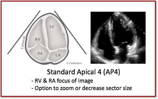
The ASE suggests measuring the RA size in the Standard AP4 window. This view allows us to image the atrium at it’s widest diameter, at end-systole.

Standard Apical 4 Chamber (AP4)


Standard AP4

Right Atrium

The ASE explains two methods for measuring the size of the RA:

  1. 2D Volume Measurement
  2. Linear Measurements

The recommendation for quantifying the size of the right atrium is to measure the right atrial volume.  The benefits to performing RA volume over linear measurements is:

  • More representation of the actual RA size
  • More robust and accurate values compared to linear measurements

In the case where you do not have full visualization to perform RA volume, then linear measurements are a good alternative method.

HOW TO PERFORM CORRECT RA VOLUME MEASUREMENT

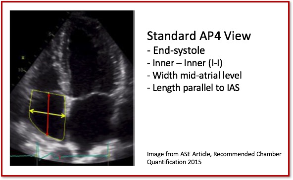
This measurement is performed from a single-plane image using either the area-length or disk summation technique.  Select which method (calculation) you will use on  your ultrasound system.  We personally prefer the disk summation technique.  With either method:

  • Standard AP4 view
  • End-Systole at largest diameter
  • Exclude tracing areas between leaflets, IVC, SVC and RA appendage
  • Area-length
    • Area
      • Trace the right atrium from the level of tricuspid valve annulus
      • Connect two annulus sides with a straight line across
    •  Length
      • Length is perpendicular to annular plane
  • Disk Summation
    • Trace the right atrium from the level of tricuspid valve annulus
    • The software package will connect the two annulus sides with a straight line
    • Remember the length MUST be perpendicular to the straight line connecting the two annulus sides.  This is important for accurate disk summation calculations.


Right Atrium Volume Measurement

RA Volume Reference Ranges

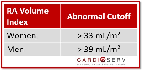
With the updated ASE guidelines, they explained that gender plays a key role when determining the abnormal values of the RA size.  Remember the referenced values provided for RA volume are:

  • Gender specific
  • Indexed to BSA

Right Atrium Volume Values

2D LINEAR RA MEASUREMENTS

As we all know that the heart is not a single-dimension structure.  Research and technological advancement in ultrasound has allowed echo to move from linear measurements to preferred volumetric methods of evaluating heart structures. With that said, if you cannot fully see the RA border properly trace than linear measurements are the next option for providing a measurement.

The updated chamber quantification guidelines provide the two linear measurements to perform, if you are unable to obtain a RA volume.

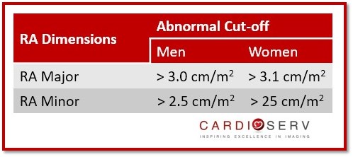
The two measurements to obtain are the:

  1. RA Minor Axis (width)
  2. RA Major Axis (length)

How to perform:

  • Standard AP4 view
  • End-Systole at largest diameter
  • RA Minor Axis (width):
    • Distance width from lateral wall to intra-atrial septum (IAS)
    • Mid-atrial level
    • Inner-edge to inner-edge (I-I)
  • RA Major Axis (length):
    • Length from center of valve annulus to center of superior RA wall
    • Parallel to intra-atrial septum (IAS)
    • Inner-edge to inner-edge (I-I)

Right Atrium Linear Measurement

Remember…Even the linear measurements are indexed to BSA!

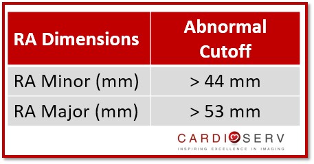
The ASE updated RA dimension ranges are now gender specific and indexed to BSA.  This is an update from the previous reference ranges last published in 2010 as the prior guidelines did not have enough data to provide gender specific guidelines and the linear dimensions previously provided were not indexed to BSA.

Right Atrium Linear Dimensions BSA

IF your laboratory has older machines and/or do not have the ability to index these linear measurements, as a guide you may choose to use the 2010 ASE RV Guidelines for linear measurement reference values.  Here is an easy reference chart to go by in your scanning laboratory:

Right Atrium Linear Measurements

We have had success manually adding indexed volume measurements to US systems for some of our clients.  We suggest that you reach out to your ultrasound vendor for help creating an indexed RA volume if this measurement is not readily available.

RA/RV measurements and cardiac cycle timing

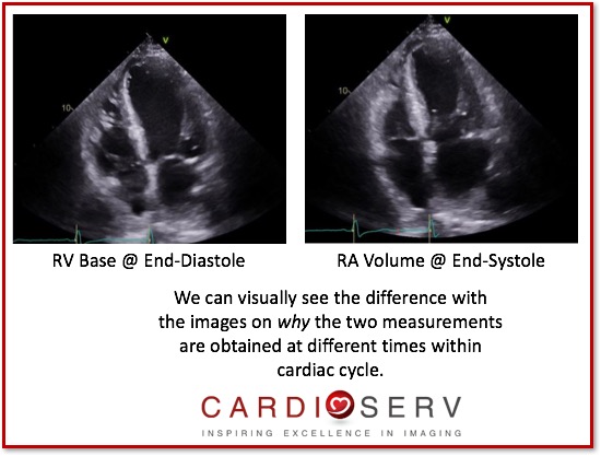
It is important that when we are performing the methods to quantify the size of the RV and RA, that we play attention to the correct timing of the cardiac cycle when we measure. Remember for the RV, we need to measure at end-diastole (when the RV is at its widest width). When obtaining the volume for RA, it is at end-systole (when the RA has the largest volume).

Diastole vs Systole

10 Tips for Correct RA Size Quantification

  1. RA Volume is the recommended method
  2. RA measurements are indexed to BSA and gender!
  3. Make sure equipment is indexing volume to BSA
  4. Use the standard apical 4 view
  5. Measure at end-systole (largest atrium diameter)
  6. Inner – to -inner edge (I-I)
  7. If you cannot see it, do NOT measure it!
  8. If you cannot trace the RA use linear dimensions
  9. Preferred method for reporting RA linear measurements are gender specific and indexed to BSA
  10. Ask your ultrasound vendor for help if you are having a hard time finding these calculations in your US system!

Conclusion

ASE provides great information on how to properly evaluate the ‘forgotten right heart’.  Here at CardioServ, we want to help provide you with easy tips to correct quantification techniques.  Our goal is for you to add right heart quantification to your routine scanning protocol.   Next week, will go further into recommend methods on how to quantify the systolic function of the RV via TAPSE and S’ Wave. We love your comments, feedback and questions! Please keep them coming and stay connected with us on Facebook, Twitter and LinkedIn!

Want a CME for reading this blog? Click below to access CardioServ’s online CME platform offering Cat. 1 AMA CMEs!

Access CMEs


Andrea Fields MHA, RDCS, Cardiac Clinical Director

Connect with me on LinkedIn


References

Lang, R. M., MD, Badano, L. P., MD, & Mor-Avi, V., PhD. (2015). Recommendations for Cardiac Chamber Quantification by Echocardiography in Adults: An Update from the American Society of Echocardiography and the European Association of Cardiovascular Imaging. JASE, 28(1), 1-53. Retrieved March 1, 2017, from http://asecho.org/wordpress/wp-content/uploads/2015/01/ChamberQuantification2015.pdf


adult echoASE guidelinescardiac chamber quantificationcmeechoechocardiographyRARA MeasurementRA Volumeright atriumRight Hearttechnical measure

Our Services

LET US KNOW WHAT YOU THINK...

Meena Sumathy
Excellent article. Thanks for sharing.
Emmeretta Pippen
Excellent
Vicky
This is very helpful
Rhonda Hassam
"Standard AP4 view" in "how to perform correct RA volume measure" looks like a focussed RV/RA view. Is this correct?
Cardioserv
The RA volume is measured in a standard apical 4 view. As you are focusing only on the RA it is OK to narrow your sector to show just the right heart.
Scott
What is the formula for RAVI so that I can enter into our calculation package? Thanks!
Cardioserv
It is a very long complicated formula (especially if using the disc summation formula). We suggest that you check to see if the formula already exists and just needs to be "turned on". If not your best bet is to reach out to the vendor as they will either modify the LA volume you already have within your system or create a new formula from scratch.
Cardioserv
It can easily be custom built if the RA volume formula already exists on your machine. (Start by checking to see if you need to "turn on" the RA volume measurement). To customize the RA volume formula, add a copy of the existing RA volume formula, re-name the formula to RAVI (on a GE - double click on the = sign, which opens an area where custom formulas can be added). Then add RA volume/BSA.
Dr Gautam Sharma
Quite detailed explanation...Keep it up
Davood Moayed
Thank you, nice presentation for RA measurements.
Alessandro Bonzano
Excellent and useful
Krishnan
Excellent review

Your email will not be displayed.

Required *

Blog
Sign Up

Accreditation
services

Accreditation Help
Online Echo CMEs
Echo Accreditation Software
Free
Consultation

Search
for

Blog
Categories

Blog
Archive

Tags
Used

Show tags
Need Help? Share