Written by Judith Buckland MBA, RDCS, FASE
Medical Ultrasound Awareness Month!
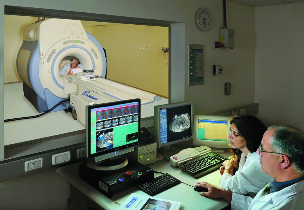
Medical Ultrasound Awareness Month (MUAM) is held annually in October to create awareness of the role diagnostic medical sonographers play in the medical community and to educate the public about medical ultrasound and its many uses in healthcare....
Written by Judith Buckland MBA, RDCS, FASE and Andrea Fields MHA, RDCS
In the past few weeks, we have reviewed different quantitative methods for evaluating the right ventricular function with echocardiography to include:
TAPSE
S’ Wave
FAC
This week we will review how to use the RIMP method to assess RV function. You will learn 4 concepts regarding...
Written by Judith Buckland, MBA, RDCS, FASE and Andrea Fields, MHA, RDCS
Last week, we talked about two methods for quantifying the function of the RV (TAPSE & S’ Wave). We selected these methods to explain first due to ease, reproducibility and established prognostic value. As some of our readers pointed out and as we discussed the TAPSE and S' Wave methods...
Written by Angela Mills, BA, RVS
What’s wrong with this vascular image?
Modality: Carotid Duplex
View: Long axis CCA
Answer:
This image demonstrates prox CCA that was labeled and measured as a distal CCA.
Correct Technique:
The Proximal CCA placement should be very low on the neck usually close to the collar bone
The...
Written by Judith Buckland
Here is a great case from Dr. Wharton on Medscape of a 45 year old patient with hyperlipidemia (high LDL-C and high triglycerides) taking a high-potency statin who presents with pericardial effusion and signs of tamponade.
What should be done next?
a) CT/MRI
b) Pericardiocentesis
c) Liver...