Echo, Vascular and Nuclear
Accreditation and Education

Author name: Don Gerig, RDCS

Sierra Hendren graduation

A Student Sonographer’s Story: Perseverance, Purpose, and the Power of Support

Every so often, we receive a message that reminds us why we do what we do. Recently, we received an email from Sierra Hendren, a cardiovascular sonography graduate from the Class of 2026. Her message was more than a thank-you note. It was a reminder of the dedication, resilience, and heart that new sonographers bring […]

A Student Sonographer’s Story: Perseverance, Purpose, and the Power of Support Read More »
ultrasound sonographer shortage

Growing Demand, Shrinking Workforce: The Sonographer Shortage Facing Imaging Labs

Across the United States, and around the world, one issue is becoming increasingly difficult for imaging labs to ignore: the shortage of qualified sonographers. While many departments have felt this operationally for years, recent workforce data now confirms what labs are experiencing every day: persistent staffing shortages, rising vacancy rates, increasing workloads, and growing pressure

Growing Demand, Shrinking Workforce: The Sonographer Shortage Facing Imaging Labs Read More »

goal-directed valsalva device for HCM LVOT obstruction echocardiography

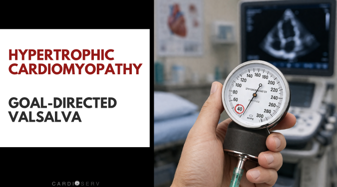
Goal-Directed Valsalva in HCM: How to Unmask LVOT Obstruction in the Echo Lab

In part 1 of our hypertrophic cardiomyopathy (HCM) series, we reviewed the key echocardiographic clues that should raise suspicion for hypertrophic cardiomyopathy. In part 2, we focused on how to differentiate LVOT obstruction flow from mitral regurgitation. Now, in this third and final article, we’ll cover one of the most important ways to unmask obstruction

Goal-Directed Valsalva in HCM: How to Unmask LVOT Obstruction in the Echo Lab Read More »

differentiate LVOT from MR Doppler in hcm

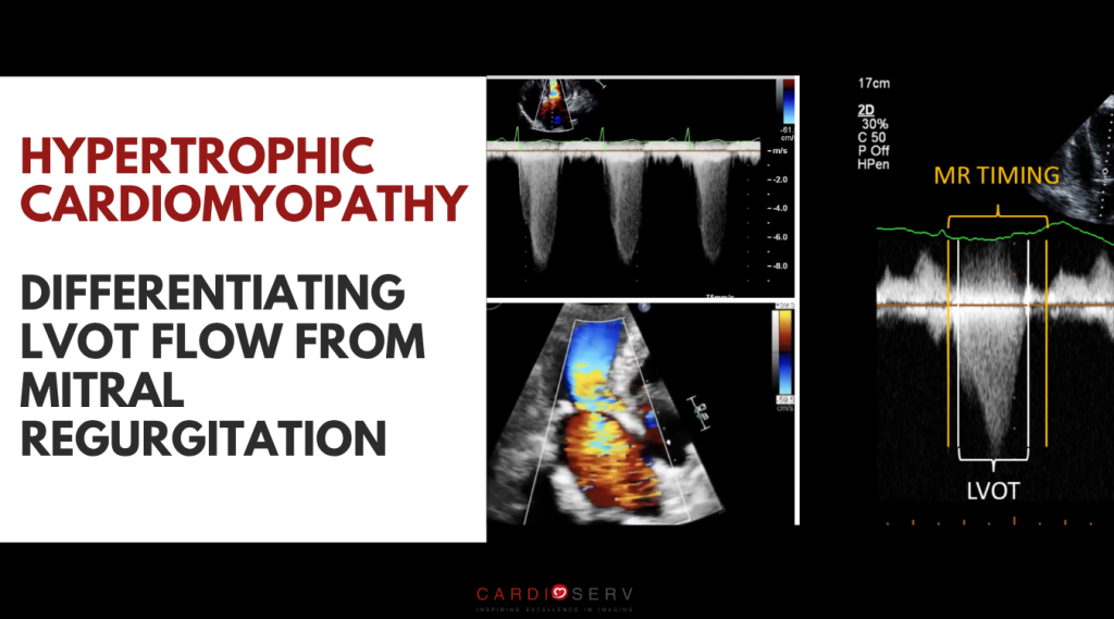
LVOT vs Mitral Regurgitation in HCM: How to Tell the Difference on Echo

In part one of our HCM series you learned key echocardiographic clues that should raise suspicion for hypertrophic cardiomyopathy.  In this article we’ll look at one of the more challenging aspects of scanning patients with obstructive hypertrophic cardiomyopathy: accurately differentiating LVOT vs Mitral Regurgitation in HCM. Both produce high-velocity systolic jets and sometimes even overlap

LVOT vs Mitral Regurgitation in HCM: How to Tell the Difference on Echo Read More »

hcm on echocardiogram

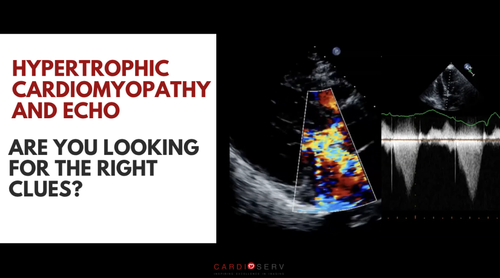
Hypertrophic Cardiomyopathy in Echocardiography: What You Might Be Missing

Hypertrophic cardiomyopathy (HCM) is more common than most sonographers realize, and it’s missed more often than anyone would like to admit. In fact, according to ASE, HCM may affect as many as 1 in 200 patients, yet a large percentage remain undiagnosed for years. Many of these patients are already being scanned in everyday echo

Hypertrophic Cardiomyopathy in Echocardiography: What You Might Be Missing Read More »

echoworkflows

EchoWorkflows: A Practical Tool for Applying ASE Guidelines in a Busy Echo Lab

CardiServ is committed to supporting individuals and organizations that share our values of inspiring excellence in echocardiography.  I recently had the opportunity to interview Michael Owen, RDCS, RVT, a sonographer and the developer of EchoWorkflows.com. I also had the chance to put his tool to the test in a real-world clinical environment in a busy

EchoWorkflows: A Practical Tool for Applying ASE Guidelines in a Busy Echo Lab Read More »

constrictive pericarditis vs restrictive cardiomyopathy

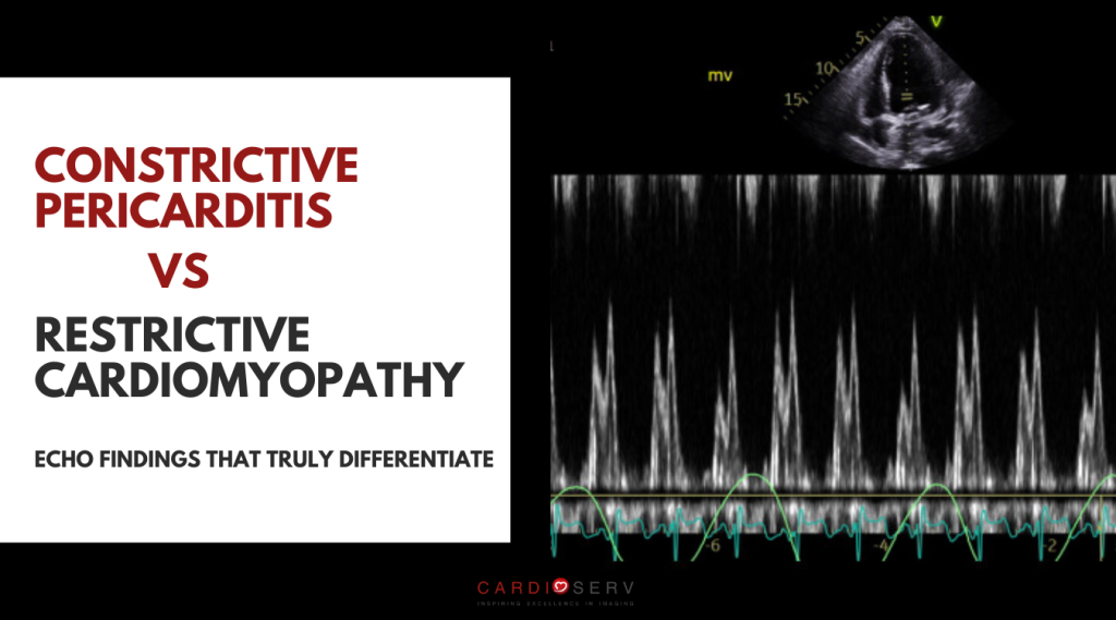
Constrictive Pericarditis vs Restrictive Cardiomyopathy – Echo Findings That Truly Differentiate

Both constrictive pericarditis and restrictive cardiomyopathy limit ventricular filling, but for completely different reasons.

They share nearly identical symptoms and often similar Doppler findings such as dyspnea, edema, JVD, preserved EF and abnormal filling, which is why they’re so commonly mistaken for one another.

But they arise from completely different mechanics, one from the outside of the heart, one from the inside.

Constrictive Pericarditis vs Restrictive Cardiomyopathy – Echo Findings That Truly Differentiate Read More »

pericardial effusion mimics

How to Recognize a Pericardial Effusion on Echo: and Avoid Common Mimics

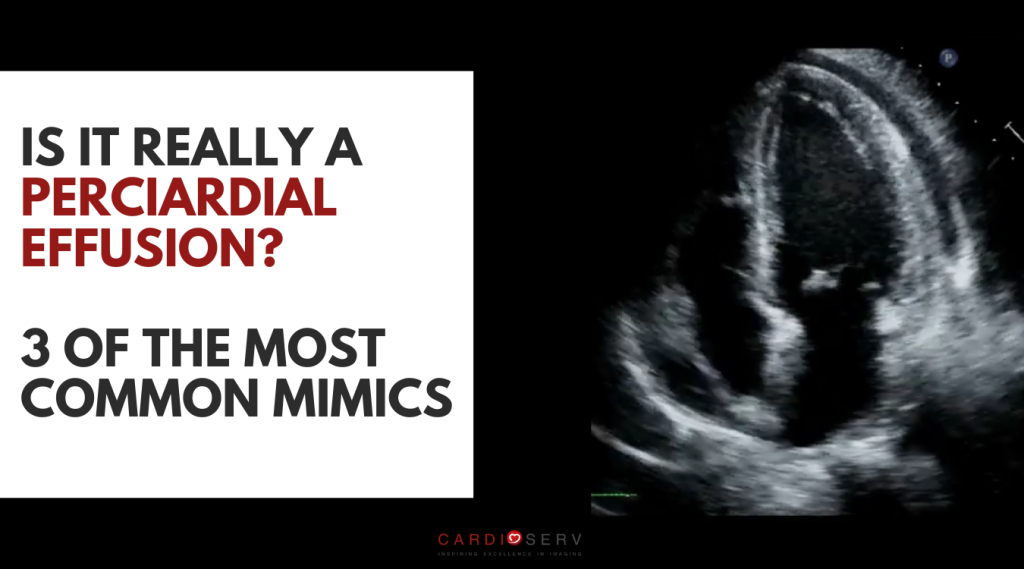
After learning the pericardial disease spectrum in Part 1 of our pericardial disease series, the natural next step is mastering the first question in the Three-Step Framework: Is there fluid? Seems like a simple enough thing to do, right? Well, it’s not always as straightforward as you might hope.  Misinterpreting an effusion can lead to

How to Recognize a Pericardial Effusion on Echo: and Avoid Common Mimics Read More »

spectrum of pericardial disease

Understanding the Spectrum of Pericardial Disease: From Effusion to Constriction

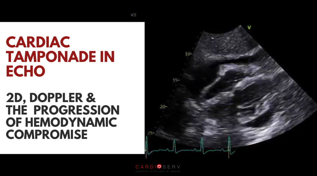
While we often think of the pericardium as a protective sac, it’s so much more than that. Under normal conditions, it moves and stretches with every heartbeat, quietly accommodating changes in volume and pressure. 

But when it loses that flexibility, the pericardium can transmit pressure back to the heart or even restrict its ability to fill. 

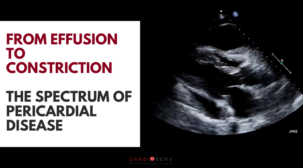
And that’s where the pericardial spectrum begins. A continuum that ranges from simple effusion to hemodynamic tamponade and, in chronic or recurrent inflammation, to constriction. Understanding this progression changes the way we view the pericardium and how we interpret our echo findings.

Understanding the Spectrum of Pericardial Disease: From Effusion to Constriction Read More »

Scroll to Top