Last Updated on January 30, 2024 by Hannes van der Merwe
ASE recently released a new guideline paper for contrast echo. First you will notice by the title of the paper that ASE is now using Ultrasonic Enhancing Agents (UEA), or ‘enhanced echo’ to replace the confusing term “contrast” echo. Thank goodness…no more stories from patients about how they are allergic to sea food and CT contrast!
To be honest, I am not usually a big fan of the topic of contrast…one too many contrast meetings back in the early days! I hesitantly began reading the newly released guideline paper and to my pleasant surprise, I found lots of useful nuggets of information worth sharing. This week we will review clinical application updates related to LV volume and EF with the use of Ultrasonic Enhanced Agents. These updated guidelines were released after extensive research conducted since the last 2008 ASE consensus paper.
LV VOLUME QUANTIFICATION WITH UEA (CONTRAST)
- End diastolic volumes measured with UEA (get use to not calling it contrast!) are significantly larger than studies performed without enhanced echo AND correlate better with cardiac MRI.
- To account for the larger size using UEA (contrast), the writing group proposed new values with emphasis on the need for additional research.
WHY IS VOLUME SIZE LARGER WHEN USING UEA? (CONTRAST)
- LV volume measurements are performed via Simpson’s bi-plane method. Read how to perform here!
- This requires tracing the blood-tissue interface, specifically the interface of the compacted myocardium and LV cavity.
- We know that artifacts, noise, trabeculations, etc. can make bi-plane volumes challenging to accurately measure, but what other reasons affect why LV volumes are significantly larger with the use of UEA?
- ASE explaines how when using UEA (contrast), the opacified blood “fills the space among the LV trabeculations up to the compacted myocardium, allowing more accurate and reproducible tracings to be performed”.
- The filling of this ‘space’ is what allows for a more accurate measurement and creates a larger cavity tracing.
LV EJECTION FRACTION USING UEA (CONTRAST)
It is easy to get caught up in the subjective nature of ejection fraction assessment. If you have ever attended an echo QI meeting, the peer review session can seem monotonous and sometimes even pointless. Please remember that for certain patients, both the accurateness and reproducibility of the LV EF is a matter of life and death, or at least quality of life. Think about the patients that are being considered for a defibrillator or life-vest, or the many cancer patients receiving chemo that need serial repeat echocardiograms to assess for cardiotoxicity. Getting the EF right is relevant!
The latest research shows that when using ultrasonic enhancing agents (contrast) during echo:
- Accurate EF assessment via echo compared with cardiac MRI improves
- Inter-observer variability is significantly reduced
ASE Recommendation: “Ultrasound enhancement should be used in all patients in whom quantitative assessment of LV EF is important to prognosis or management of the clinical condition.”
SUMMARY
ASE released updated guidelines for UEA (contrast echo) and this week we reviewed how these updates relate to LV volume quantification and ejection fraction assessment.
3 TAKE-AWAYS:
- Ultrasound Enhancing Agents (UEA) and/or ‘enhanced echo’ is replacing the old terminology of ‘contrast echo’.
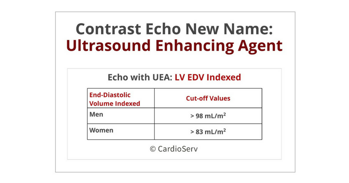
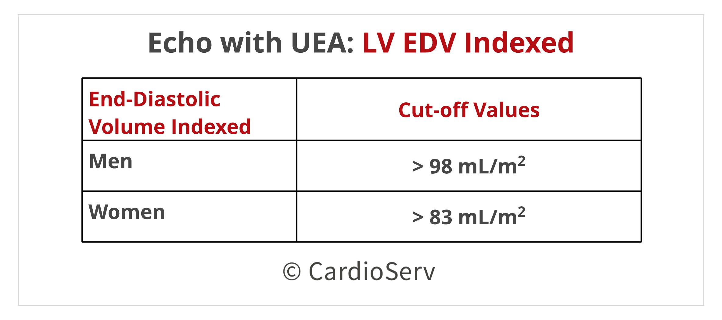
- Enhanced echo LV volumes are larger than volumes obtained though echocardiograms without UEA. The new proposed cut off range is:
- Women: 83 mL/m2
- Men: 98 mL/m2
- In situations where LV EF assessment is dependent upon patients prognosis or management, ASE recommends using Ultrasound Enhancing Agents.
Keep an eye out – we will continue to review the latest UEA guideline paper in future blogs!
Need CMEs? CardioServ now offers Cat. 1 AMA Echo CMEs!


Judith Buckland, MBA, RDCS, FASE
Stay Connected: Facebook, Twitter, Instagram, LinkedIn
Reference:
Clinical Applications of Ultrasonic Enhancing Agents in Echocardiography: 2018 American Society of Echocardiography Guidelines Update
http://asecho.org/wordpress/wp-content/uploads/2018/03/UEA-Guideline.pdf


