Last Updated on November 6, 2025 by Don Gerig, RDCS
Last week, we covered the basic anatomy of the mitral apparatus. We reviewed in detail, the various structures and their function. As we continue with our mitral regurgitation blog series, we want to touch next on, specific imaging windows to evaluate the mitral valve. Remember, when we examine the valve, we need to make sure we include all structures of the apparatus.
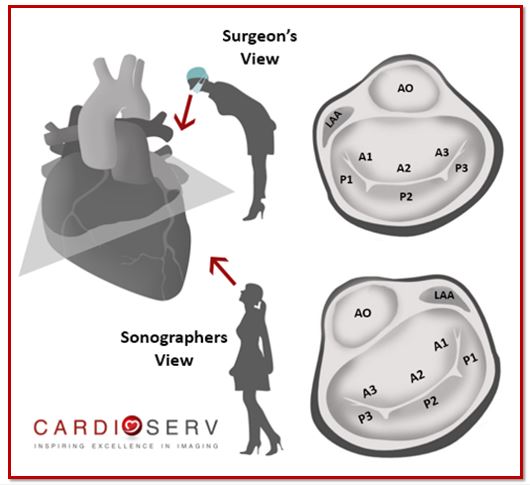
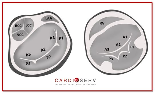
Surgeon’s View vs Echocardiographer’s View of the Mitral Valve
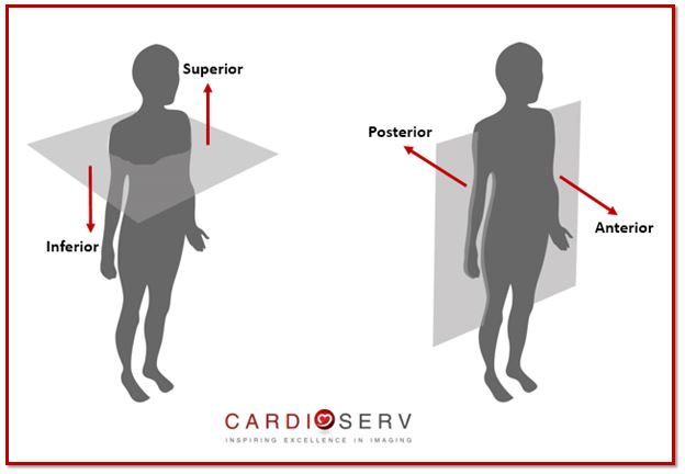
When we talk about the mitral apparatus, we need to understand how the image varies depending on the angle and viewpoint we are scanning from. As sonographers, we need to remember that echo images of the mitral valve are displayed upside down & backwards! First, let’s get our basic orientation down:
This can get a little confusing when trying to communicate with surgeons. Surgeons think of the heart in the anatomical orientation and look at the mitral valve from the left atrium. Sonographers/cardiologists, often think from an ultrasound orientation and look at the mitral valve from the left ventricle.
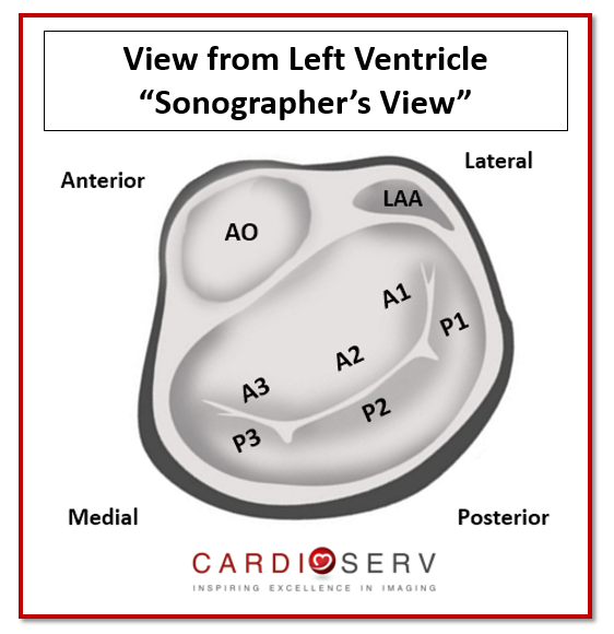
For this article, we are going to review anatomy, from the orientation of looking at the mitral valve from the left ventricle. Just as we do during a transthoracic echo. Remember, we scan upside down & backwards with the ventricles ‘on-top’ of our sector.
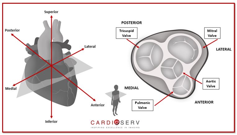
Regardless of the angle you’re viewing the mitral valve from, these anatomical tips will always help orientate you:
- A1/P1 – lateral
- A3/P3 – medial
- Aorta (AO) – anterior
- Left atrial appendage (LAA) – lateral with A1/P1
Imaging The Mitral Valve
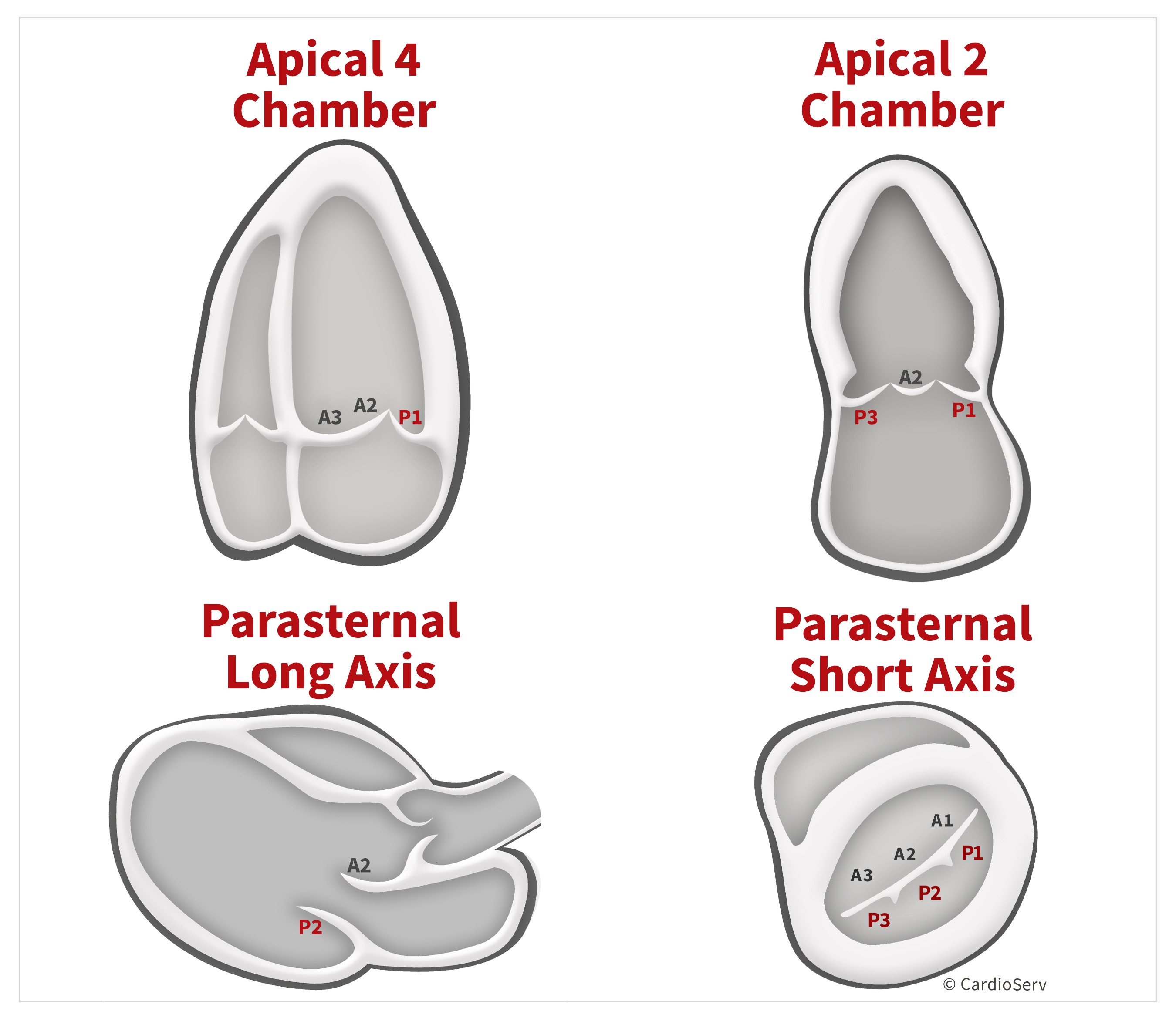
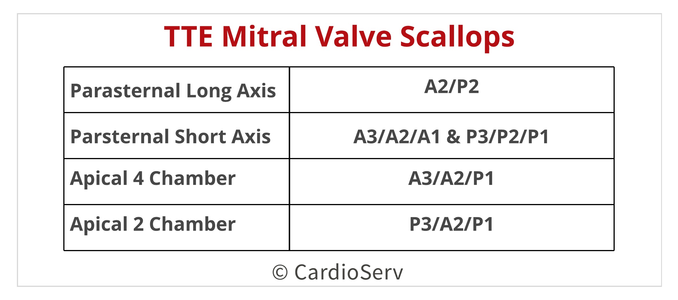
We have 4 standard views to visualize the mitral valve:
- Parasternal long axis (PLAX)
- Parasternal short axis (PSAX)
- Apical 4 Chamber (AP4)
- Apical 2 Chamber (AP2)
By angulating our transducer from our standard views, we can image more than the ‘set standard’ scallops seen. Let’s break down the four views to better explain!
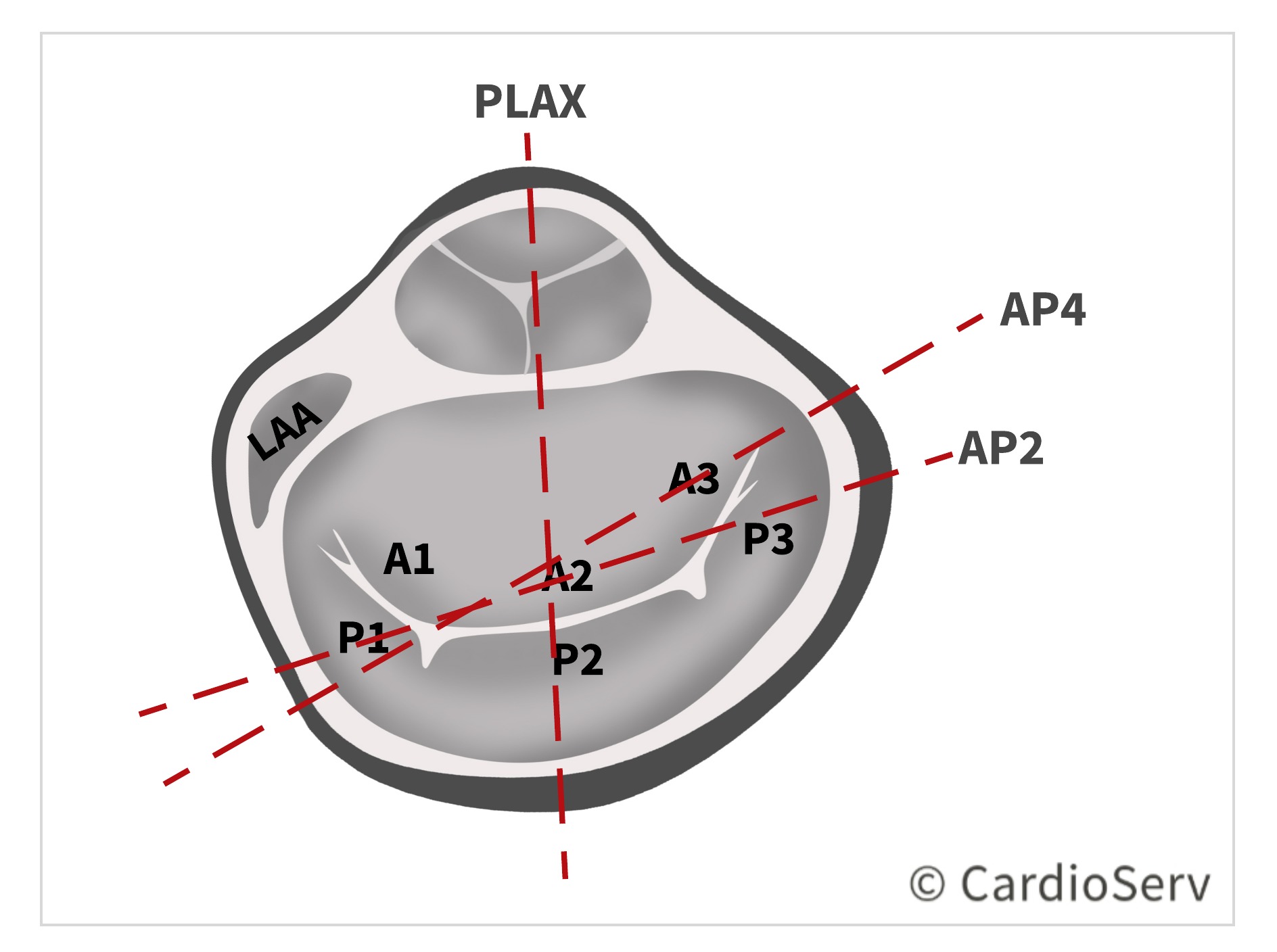
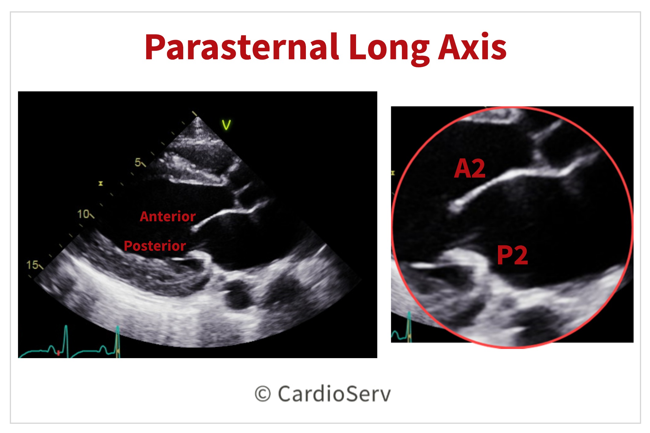
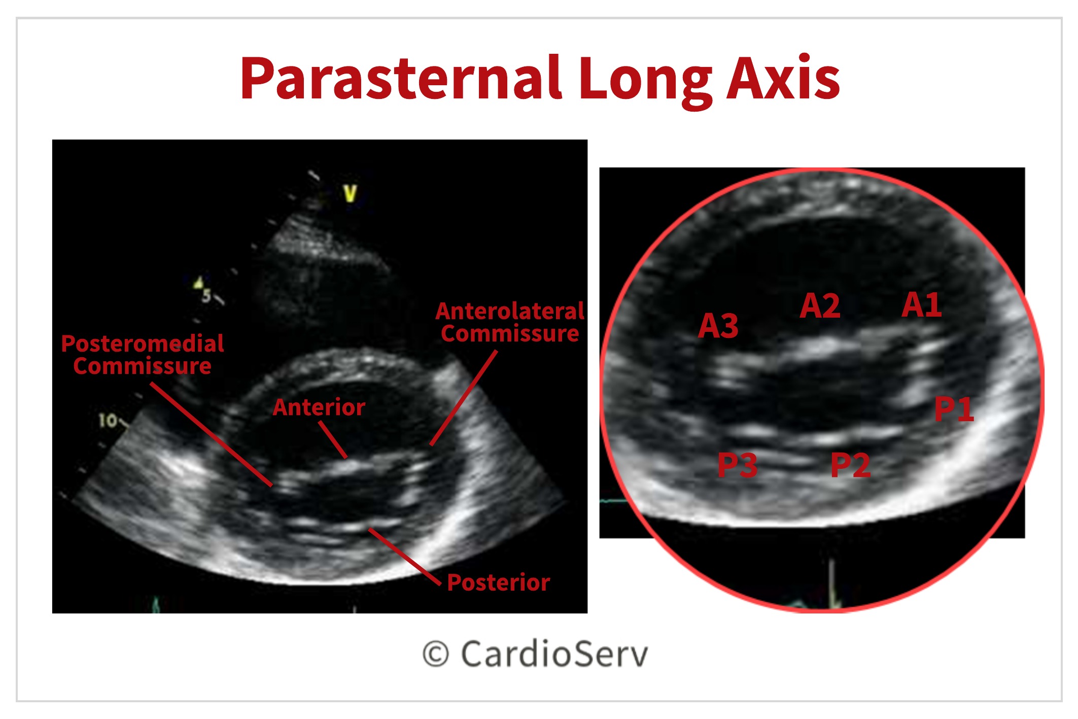
Parasternal Long Axis (PLAX)
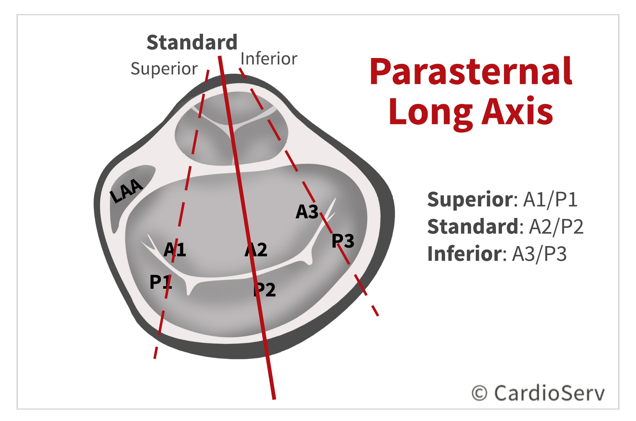
From the PLAX viewing window, we are able to see A2 & P2 of the mitral valve.
We are able to view additional scallops by a simple change in angulation of the transducer:
- Angle transducer superior (towards RV outflow) identify: A1/P1
- Angle transducer inferior (towards RV inflow) identify: A3/P3
Parasternal Short Axis (PSAX)
Mitral Valve Leaflets and Scallops:
From the PSAX window, we can visualize all 6 scallops of the anterior & posterior leaflets!
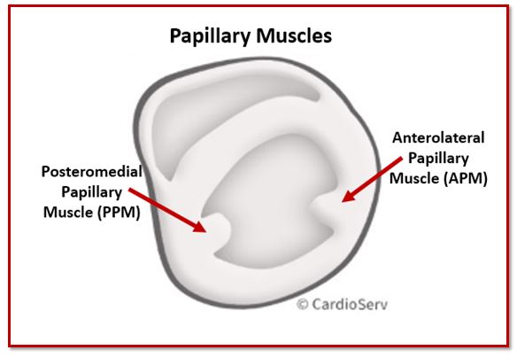
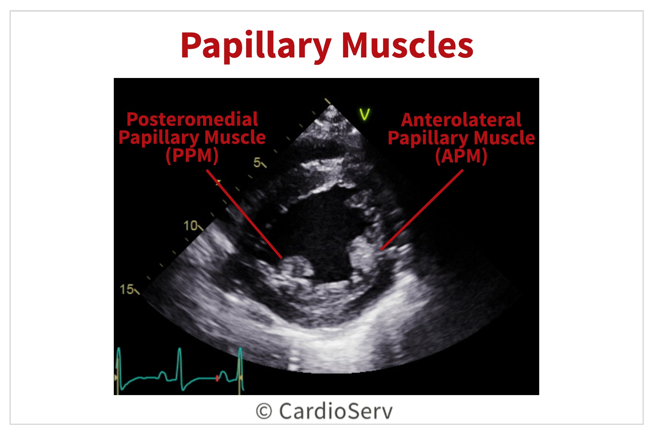
Papillary Muscles:
Apical 4 Chamber (AP4)
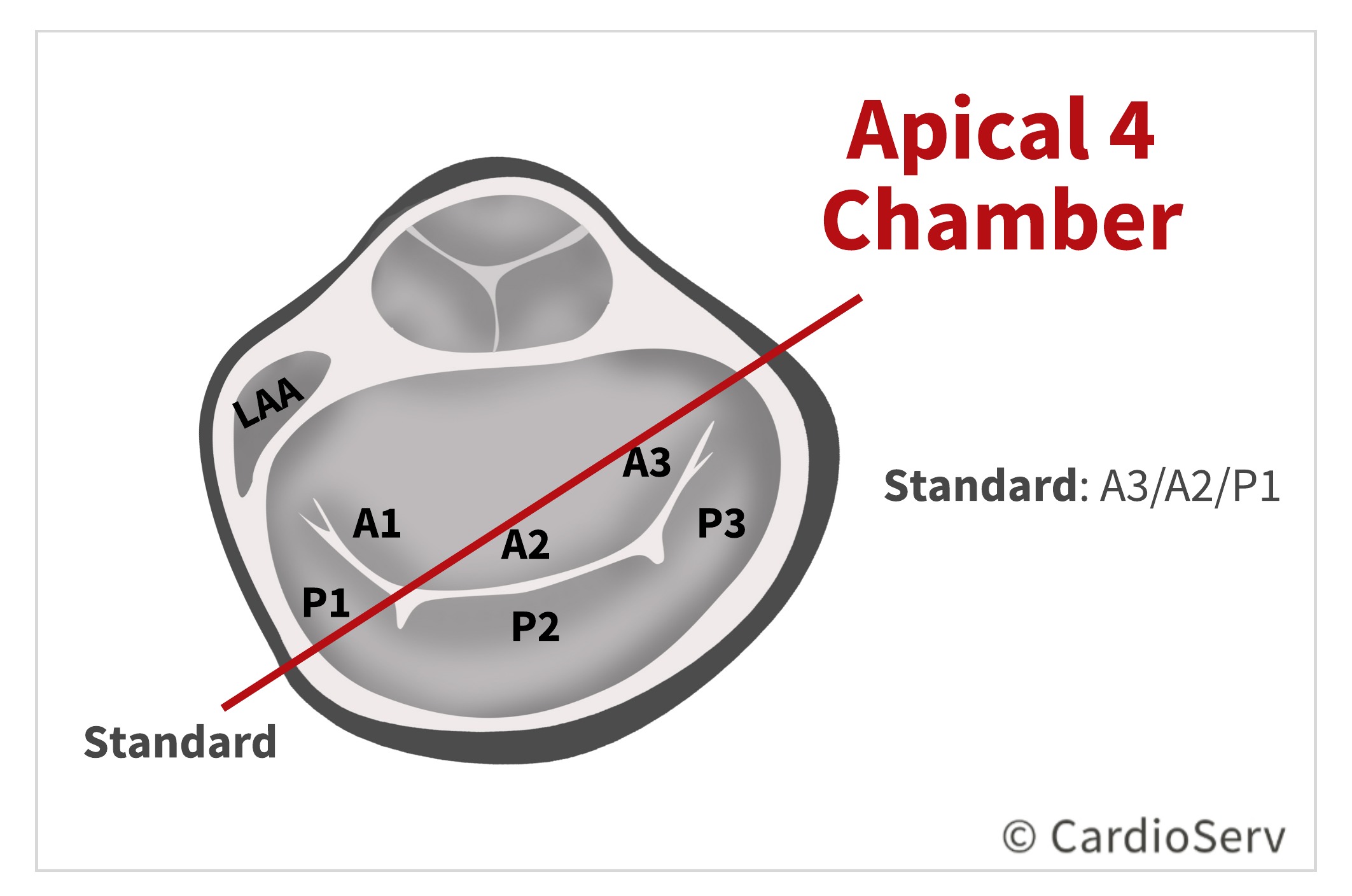
From the AP4 standard view, we can identify A3/A2/P1!
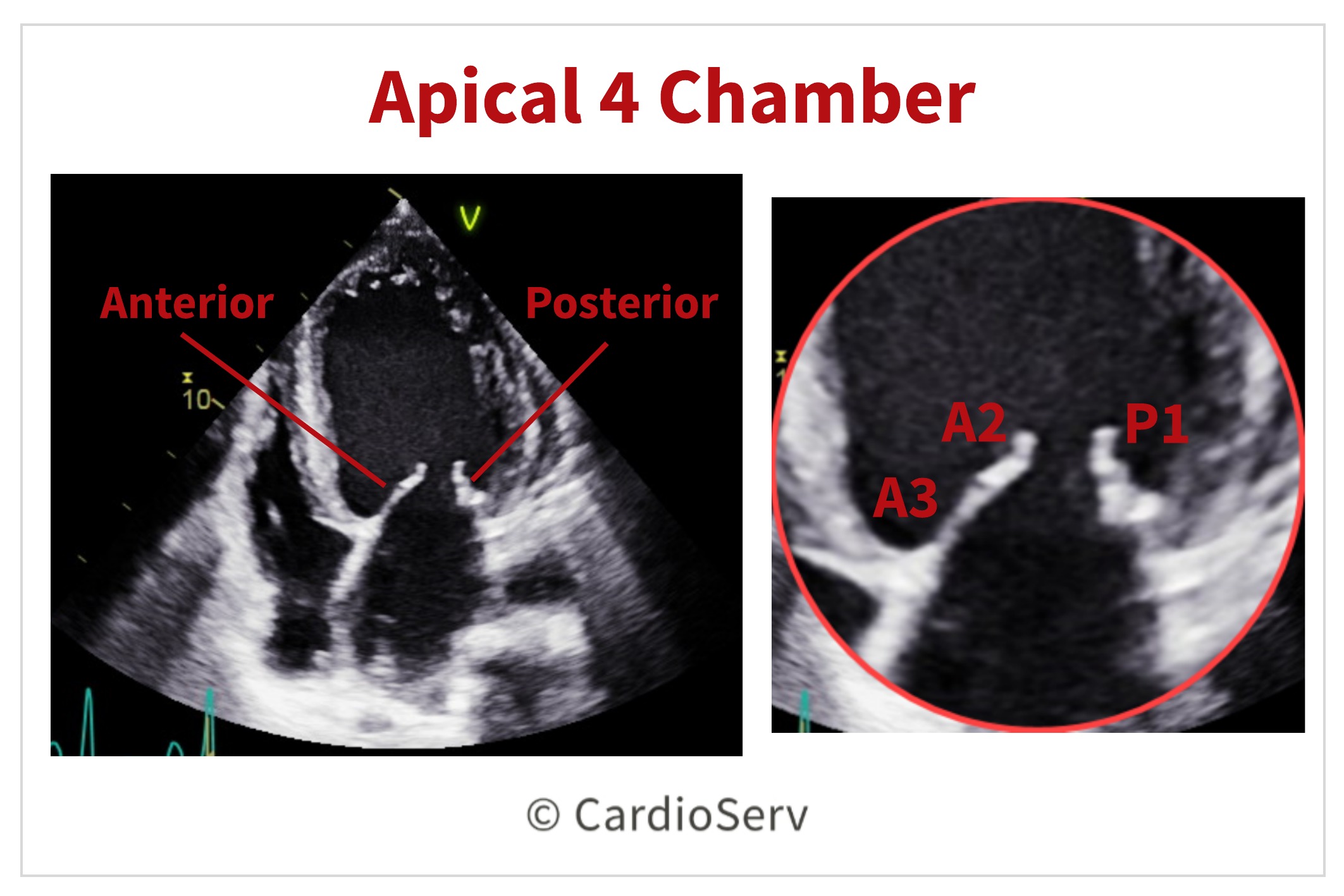
This imaging window is easy to slightly angle our transducer in order to visualize more scallops!
- Angle anterior (bringing in apical 5 chamber view) to visualize A1/P1 and the anterolateral commissure.
- Angle inferior (visualizing long-axis of coronary sinus) to visualize A3/P3 and the posteromedial commissure.
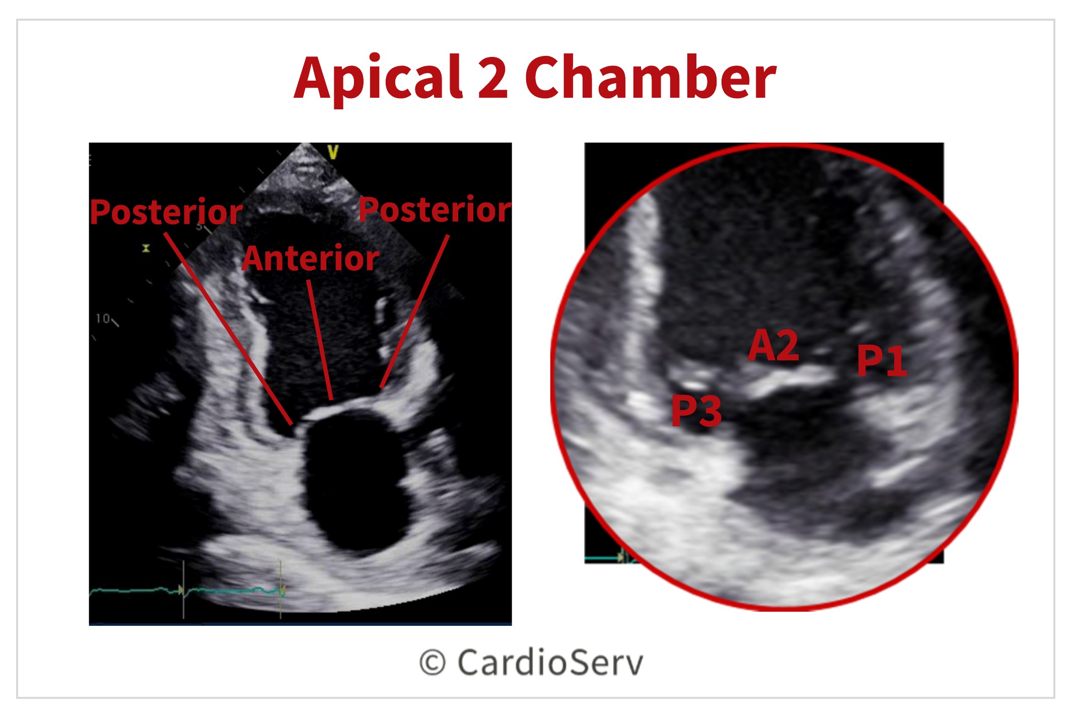
Apical 2 Chamber (AP2)
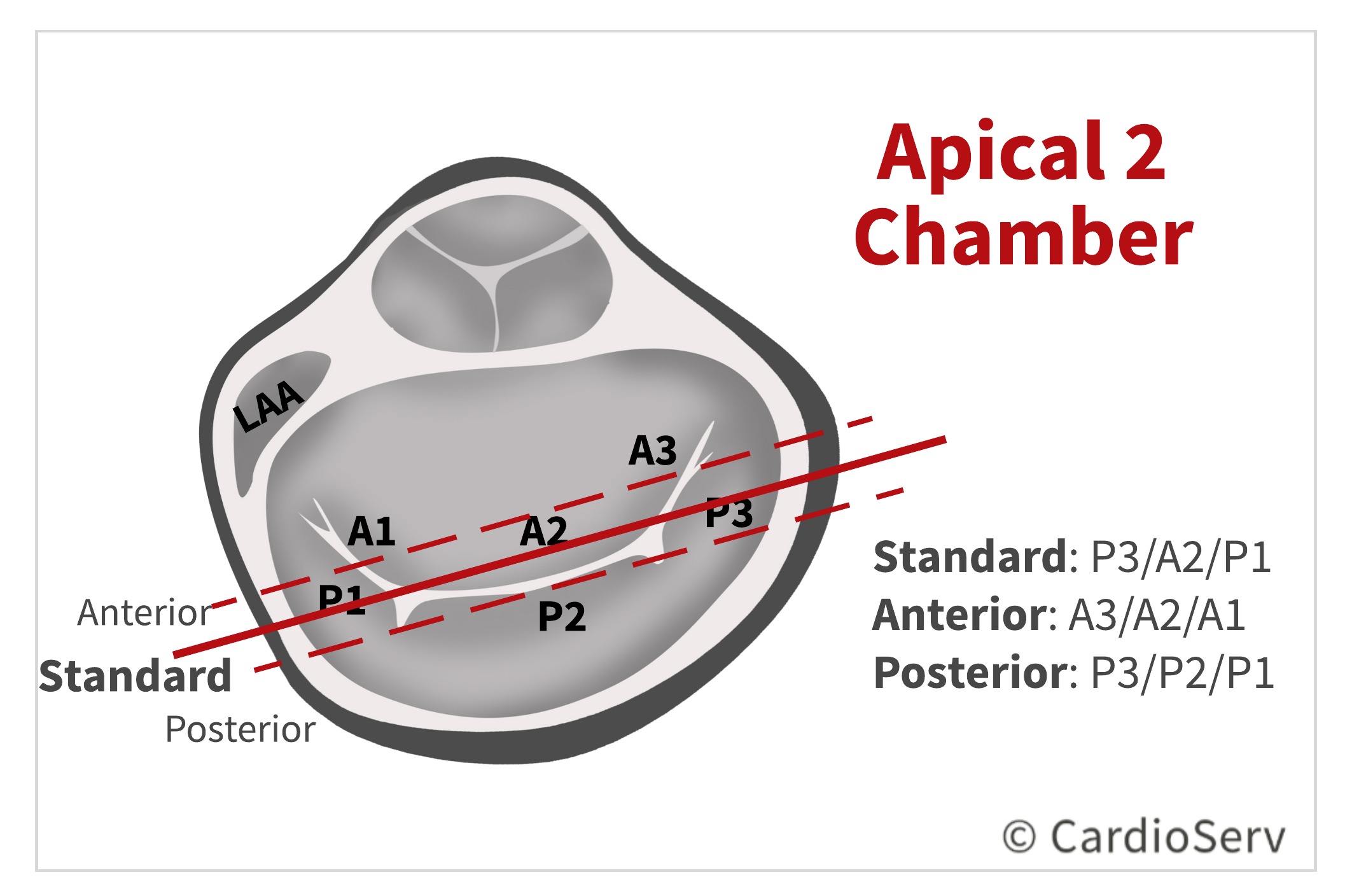
From the AP2 standard view, we can visualize P3, A2 & P1. This view can be confusing: Why do we see the middle scallop of A2? This is because we are viewing the mitral valve at an angle where both coaptation points occur, causing A2 disappears during diastole.
We can identify more scallops by angulating our transducer:
- If we angle the transducer anterior, we will visualize a longer anterior leaflet with a shorter posterior.
- If we angle posterior, we will not visualize a coaptation zone, imaging the posterior leaflet (and mostly P2).
Summary & Key Takeaways on Mitral Valve Echo Orientation
By the end of this articl, we hope that you can know the 4 standard views for imaging the mitral valve and corresponding scallops.
Remember that just because you’re imaging a specific window does not mean you are seeing the standard scallops associated with that view. The mitral scallops are small segments and can easily be viewed by a simple angulation of the transducer.
We hope you are enjoying our mitral regurgitation blog series. Now that we have our basic anatomy, structure and imaging planes covered–keep an eye out for our blog next week over mechanisms of mitral regurgitation!
Take the Guesswork Out of Mitral Valve Imaging
Mitral Valve Imaging in Echo – Visual Guide
Sharpen your MR assessment with this clear, image-rich guide to echo views, scallops, and probe positioning.
- ✔ Understand scallop anatomy in TTE & TEE with ease
- ✔ Avoid common pitfalls like foreshortened views
- ✔ Dozens of labeled diagrams for quick reference
- ✔ Practical checklists to streamline MR imaging

Andrea Fields MHA, RDCS
Stay Connected: LinkedIn, Facebook, Twitter, Instagram
References:
Prokšelj, K. (2015). Echocardiography Of The Mitral Valve. International Symposium MITRAL VALVE DISEASES IN CHILDREN AND ADULTS. doi:10.5644/pi2017.168.03
Zoghbi, W. A., MD, Adams, D., RCS, RDCS, FASE, & Bonow, R. O., MD. (2017). Recommendations for Noninvasive Evaluation of Native Valvular Regurgitation. JASE,30(4), 318-334. Retrieved June 6, 2017.
Zamorano, J. L., MD, & Badano, L. P. (2011). EAE/ASE Recommendations for the Use of Echocardiography in New Transcather Interventions for Valvular Heart Disease. JASE,24(9), 957-960. Retrieved June 6, 2017.