Last Updated on November 18, 2025 by Don Gerig, RDCS
We recently reviewed a case, during a local hospital quality improvement meeting, that required us to assess whether or not there was prosthetic aortic valve stenosis. It was interesting to touch up on the key parameters that should be assessed. This week we will review the key points for imaging and interpreting prosthetic aortic valve stenosis with echocardiography.
PROSTHETIC AORTIC VALVE STENOSIS: KEY IMAGING POINTS
Imaging the heart status post valve repair can be challenging, especially due to the artifact caused by some prosthetic valves. This is why we encourage off axis imaging, whenever necessary, to better visualize your region of interest. Pay attention to the following items during an echocardiogram of a prosthetic valve:
- Opening and closing of leaflets or occluders
- Abnormal densities (calcium, mass, vegetation)
- Stability versus rocking motion
- Aware of low flow states
- Modified views to keep artifact from regions of interest
- 3D TEE for direct viewing
- 2D remains essential due to higher temporal and spatial resolution
PROSTHETIC AORTIC VALVE STENOSIS: PRESSURE GRADIENTS AND VELOCITY
- Remember, that often the aortic valve velocity in a prosthetic valve is increased (> 2m/s), therefore velocities <3m/sec are still considered ‘normal’.
- High gradients may be seen with normal functioning valves with:
- Small size
- Increased stroke volume
- PPM (Patient Prosthetic Mismatch)
- Valve obstruction
- Mildly elevated gradient with severe LV dysfunction may still indicate stenosis
- Gradients alone are not enough to evaluate malfunctioning prosthetic valves
PROSTHETIC AORTIC VALVE STENOSIS: JET CONTOUR
The jet contour can provide us with valuable information. Although not a quantitative assessment, it is a qualitative measure and is considered a valuable index of prosthetic valve function
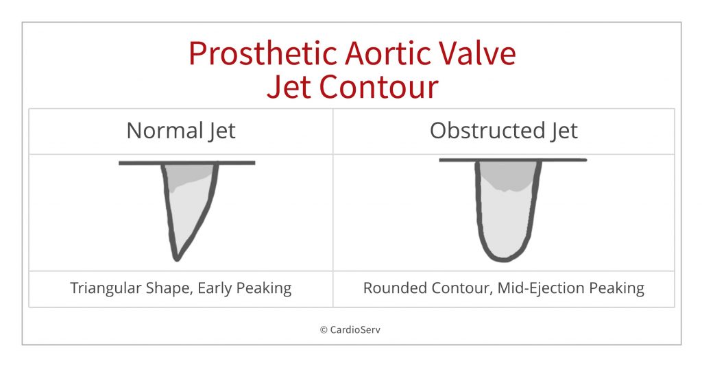
Normal prosthetic valve jet contour
- Triangular shape of the velocity
- Early peaking of the velocity
- Short acceleration time (AT)
Obstructed prosthetic valve:
- More rounded contour
- Mid-ejection peaking of velocity
- Prolonged acceleration time (AT)
- Prolonged ejection time (ET)
AV ACCELERATION TIME
While the jet contour is qualitative, we can use the acceleration time as a quantitative measure to assess prosthetic aortic valve stenosis. An acceleration time > 100 m/s differentiates between normal and stenotic prosthetic valves.
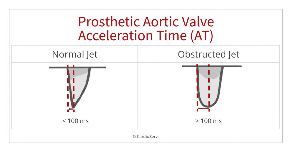
HOW TO MEASURE AORTIC VALVE ACCELERATION TIME
- CW of the AV
- Measurement from onset to peak velocity / time measurement
- >100 ms differentiates between normal and stenotic prosthetic valves
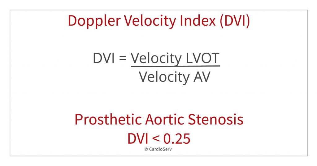
DVI – Doppler Velocity Index
Another quantitative measurement is the Doppler Velocity Index (DVI). The DVI is a dimensionless index that is great to use in the presence of prosthetic valves because the value is not flow dependent and is less dependent on valve size. It’s especially useful when the valve size is unknown. DVI < 0.25 is highly suggestive of significant valve obstruction.
How to obtain the DVI
DVI = VelocityLVOT / VelocityPrAV
- Measure max LVOT velocity in PW.
- Back away from valve just enough to avoid turbulence from the valve
- Measure the max AV velocity in CW.
- Be sure to obtain highest velocity through the valve
- Use the PEDOF probe
- Assess the AV from all windows (apical 5, suprasternal, right parasternal)
- Use the MAX velocity obtained from all views (not the average)
DVI < 0.25 is highly suggestive of significant valve obstruction
ADVANTAGES OF DVI:
- Dimensionless ratio
- Less dependent on valve size
- Useful when valve size not known
- Good when AVA/EOA cannot be obtained due to inaccurate LVOT area
- Is not flow dependent
SUMMARY
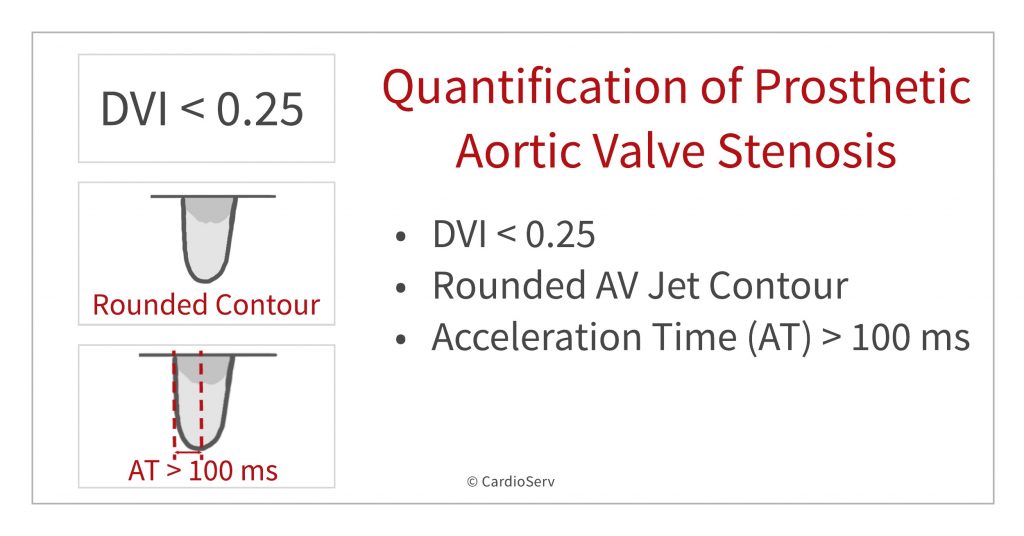
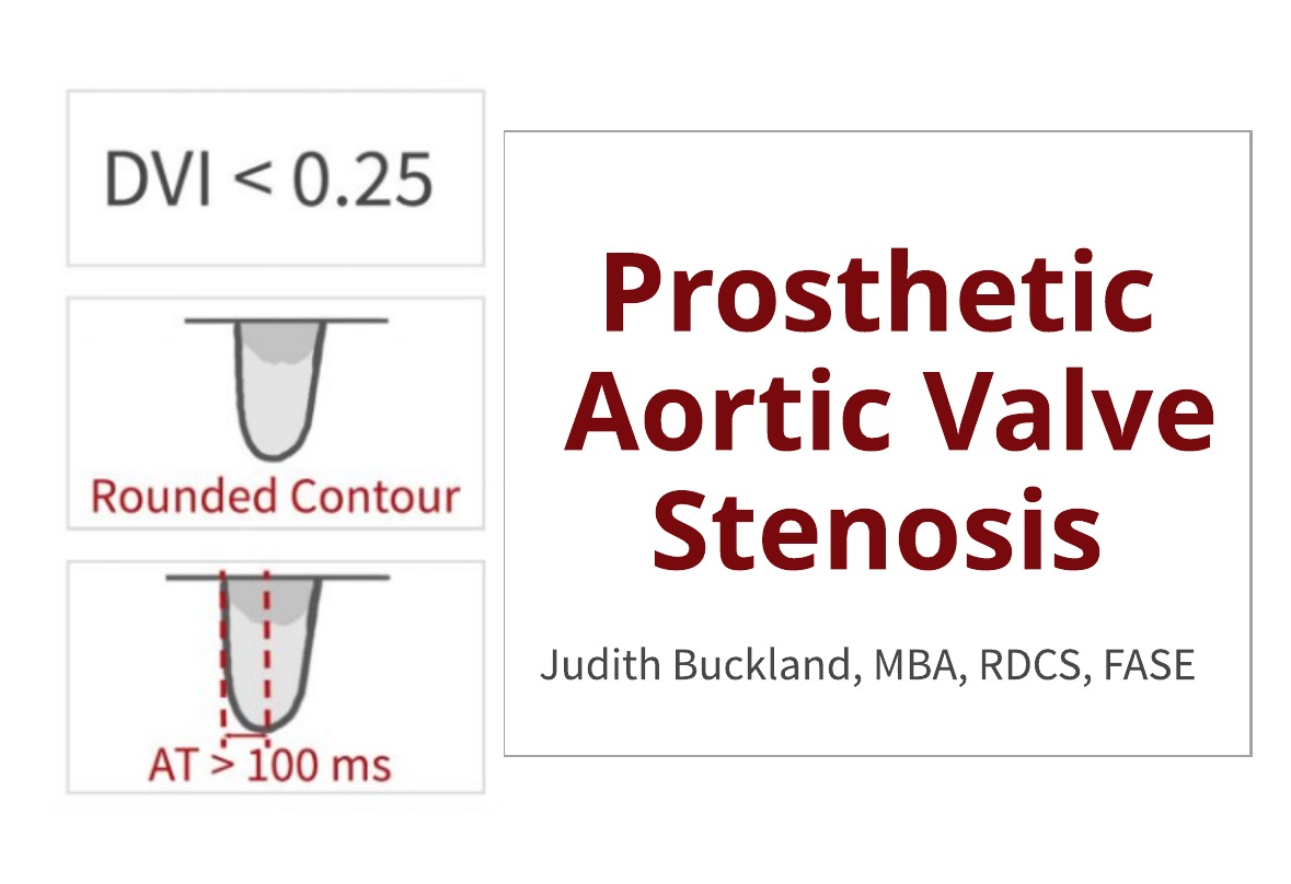
When quantifying prosthetic aortic valve stenosis on echocardiography we learned that three parameters can help us determine prosthetic aortic valve stenosis:
- DVI < 0.25
- Rounded Jet Contour
- Acceleration Time (AT) >100 ms
References
Zoghbi, et al. Evaluation of Prosthetic Valves With Echocardiography and Doppler Ultrasound. American Society of Echocardiography. 2009. Retrieved from: https://www.asecho.org/wp-content/uploads/2014/05/2009_Evaluation-of-Prosthetic-Valves.pdf

Judith Buckland, MBA, RDCS, FASE


