Last Updated on January 30, 2024 by Hannes van der Merwe
WHAT’S WRONG WITH THIS IMAGE?
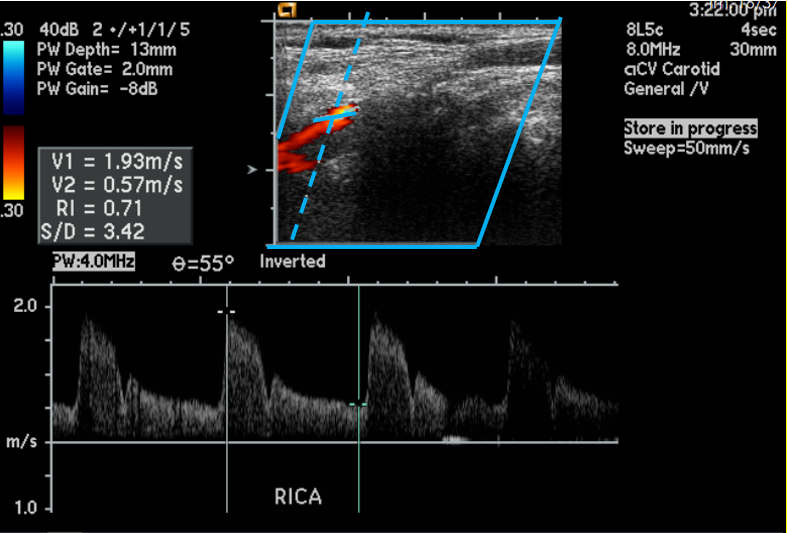
| Modality: | Carotid Vascular |
| View: | Color Right ICA Prox |
| Measurement: | RICA Prox – Peak/end diastolic velocity |

ANSWER:
- The angle is not parallel to the vessel.
- The image label is missing location (prox, mid, distal)
TIPS FOR CORRECT ANGLE PLACEMENT:
- Ensure the angle is parallel to the vessel; options include:
- Adjust the color box angle (this adjusts the Doppler beam)
- Change the angle correction to remain parallel to the vessel.
- Keep Doppler angles at < 60°
- Try and keep angles between 45° – 60°
- Occasionally, due to patient anatomy Doppler angles that are less than 45° may be encountered
- The sample gate should be centered to the flow
EXAMPLE 1:
Adjusting the color box will adjust the direction of the Doppler beam.

In this image, by adjusting the color box the direction of the Doppler beam changed. By changing the Doppler beam and correcting, the angle to display parallel to the vessel the Doppler angle changed from 55° to 60°.
EXAMPLE 2:
Changing the angle correction to remain parallel to the vessel.

In this image, the angle correction was adjusted to run parallel to the vessel wall. The angle changed from 55° to an acceptable 45°.
HOW DOES THE DOPPLER ANGLES INFLUENCE VELOCITY CHANGES?
It is drilled in us from the time we start learning in the classroom that the Doppler cursor should be aligned parallel to the vessel walls and Doppler angles should be maintained between 45°- 60° whenever possible. The Society of Vascular Ultrasound notes that occasionally, due to patient anatomy, Doppler angles that are less than 45° may be encountered and they recommend that we note this on the worksheet. Just how much does angle affect the velocity readings??
HERE ARE SOME EXAMPLES FROM INDUSTRY JOURNALS:
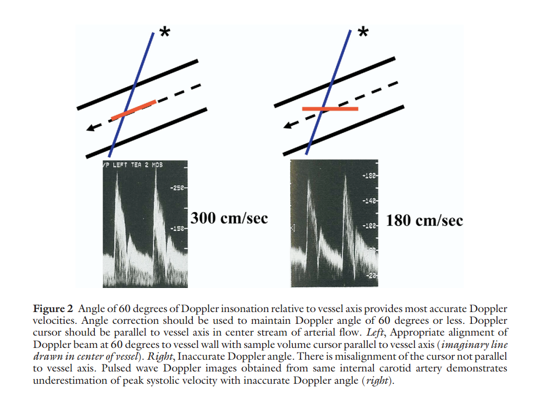
Guidelines for Noninvasive Vascular Laboratory Testing: A Report from the American Society of Echocardiography and the Society of Vascular Medicine and Biology.
- 1st image demonstrates correct placement parallel to vessel walls with an angle of 60°, which correctly measures the velocity at 300 cm/sec.
- 2nd image demonstrates incorrect placement that is NOT parallel to flow which greatly underestimates the max velocity.
SonoWorld
Below are examples from SonoWorld.com of the same vessel measured at 30° and 50°. You can see the difference in velocity readings.
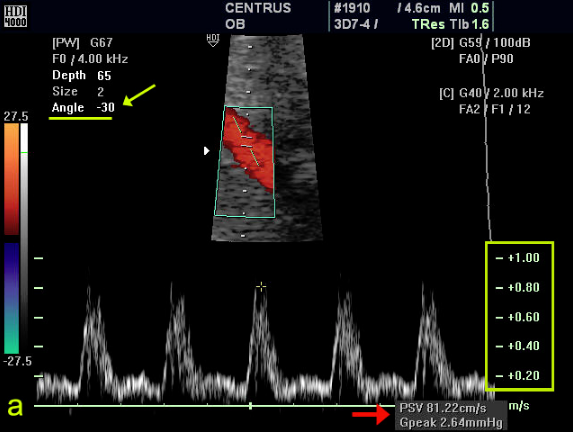 | 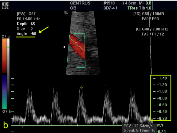 |
| Angle: 30°Velocity: 81 cm/sec | Angle: 50°Velocity: 114 cm/sec |


