Last Updated on November 18, 2025 by Don Gerig, RDCS
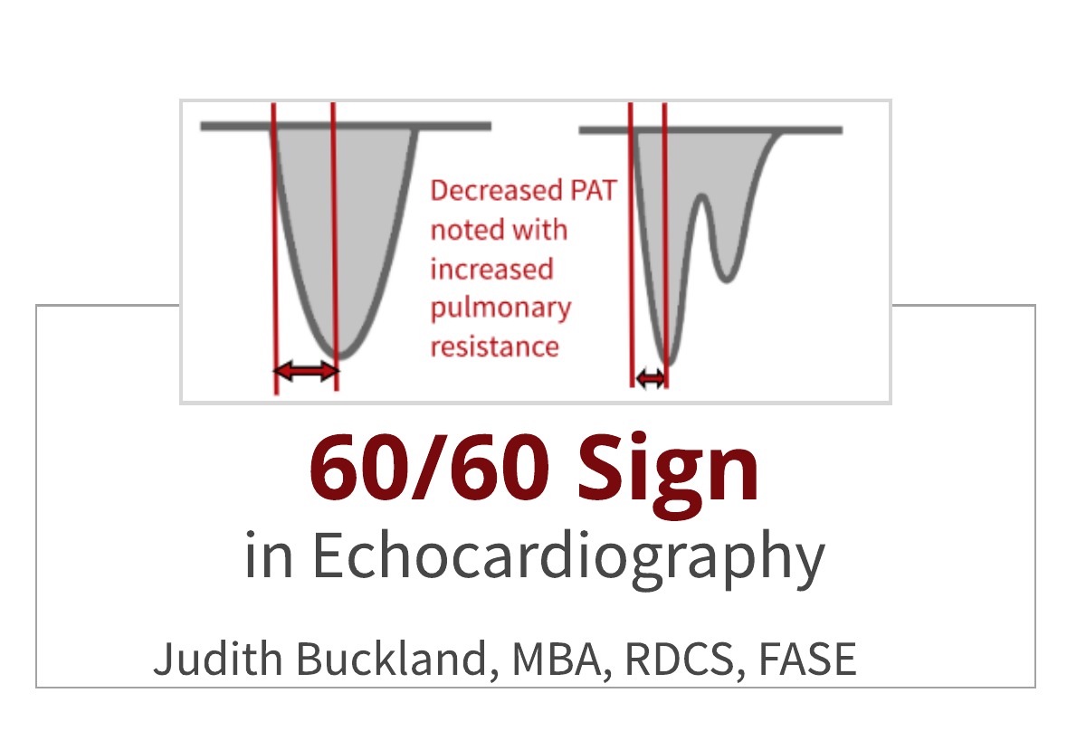
60/60 Echo Sign
Last month we presented Echo in Pulmonary Embolism. One of the echo findings associated with acute pulmonary embolism is the echo 60/60 sign. What is the echo 60/60 sign? This week we will learn the features of the 60/60 sign in echocardiography and its implications with pulmonary embolism.
WHAT IS THE 60/60 SIGN IN ECHOCARDIOGRAPHY?
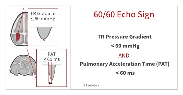
The 60/60 sign includes BOTH:
- TR pressure gradient < 60 mm Hg
- Pulmonary acceleration time (PAT) of < 60 msec
Please note that it’s the TR pressure gradient that needs to be < 60 mmHg, NOT the RVSP! I mention this because during my research I found sites, and even YouTube videos, that listed the 60/60 sign as PAT < 60 / RVSP < 60. After additional research (references below), I can confirm that the 60/60 sign involves the PAT and TR gradient. Another interesting note is that European literature uses < 60 while references in the USA use < 60.
NORMAL VERSUS ABNORMAL PULMONARY ACCELERATION TIME IN ECHO
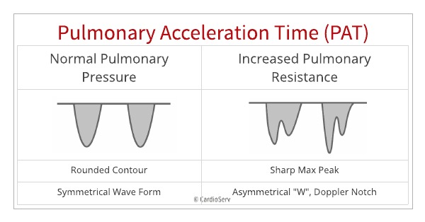
Normal PAT
- Spectral Doppler looks symmetrical
- Rounded contour
Abnormal PAT
- Sharp pointed max peak
- Spectral Doppler notch – looks like an asymmetrical ‘W’
HOW TO CORRECTLY MEASURE THE PULMONARY ACCELERATION TIME:
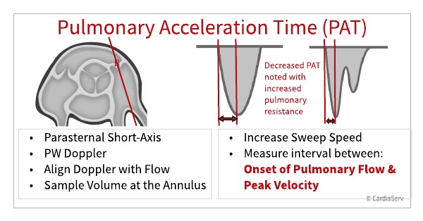
The Pulmonary Acceleration time (PAT) is the amount of time between the onset of systolic arterial flow and peak velocity. When the pulmonary resistance is high, the wave form is abnormal. The correct way to measure this is as follows:
- Parasternal short-axis view
- PW Doppler
- Align Doppler with blood flow direction
- TIP: Place sample volume at the annulus of the pulmonary valve – avoid placing the sample more proximally in the RVOT
- Use faster sweep speed (stretch out waveform)
- Measure the PAT – time interval between the onset of systolic pulmonary arterial flow (onset of ejection) and peak flow velocity
WHAT’S THE SIGNIFICANCE OF THE 60/60 SIGN?
The 60/60 sign is not sensitive for diagnosing pulmonary embolus (PE) but it is very specific (94%). Due to some controversy over the subjective nature of the McConnell’s sign, the 60/60 sign, “may be more practical in clinical practice because the criteria are more clear cut” (Lang et al, 2011).
SUMMARY
The 60/60 Sign in echocardiography is the presence of BOTH:
- TR pressure gradient < 60 mm Hg
- Pulmonary acceleration time (PAT) of < 60 msec
This sign is not very sensitive for pulmonary embolism but extremely specific. Other articles related to pulmonary embolism:
- Echo in Pulmonary Emoblism – the Clot Thickens!
- Pulmonary Embolism in Echocardiography
- TAPSE as a Prognostic Tool in Pulmonary Embolism
- Invasive Ultrasonic Procedures to Treat Pulmonary Emoblism
References
Kurzyna M, Torbicki A, Pruszczyk P. Disturbed right ventricular ejection pattern as a new Doppler echocardiographic sign of acute pulmonary embolism. The American journal of cardiology. 90(5):507-11. 2002. [pubmed]
Lang, R. M., Goldstein, S. A., Kronzon, I., & Khandheria, B. K. (2011). Dynamic echocardiography. St. Louis, MO: Saunders Elsevier.
Torbicki, A., Kurzyna, M., Ciurzynski, M., Pruszczyk, P., Pacho, R., Kuch-Wocial, A. et al.Proximal pulmonary emboli modify right ventricular ejection pattern. Eur Respir J. 1999; 13: 616–621
Kurnicka, Katarzyna et al. Echocardiographic Pattern of Acute Pulmonary Embolism: Analysis of 511 Consecutive Patients. Journal of the American Society of Echocardiography, Volume 29, Issue 9, 907 – 913
Tossavainen, E., Soderberg, S., Gronlund, C., Gonzalez, M., Henein, M., & Lindqvist, P. (2013, January 07). Pulmonary artery acceleration time in identifying pulmonary hypertension patients with raised pulmonary vascular resistance. Retrieved from https://academic.oup.com/ehjcimaging/article/14/9/890/2397468
Torbicki, A. (2005). Echocardiographic diagnosis of pulmonary embolism: A rise and fall of McConnell sign? European Journal of Echocardiography,6(1), 2-3. Retrieved June 4, 2019.

Judith Buckland, MBA, RDCS, FASE
Stay connected: Facebook, Twitter, Instagram, LinkedIn


