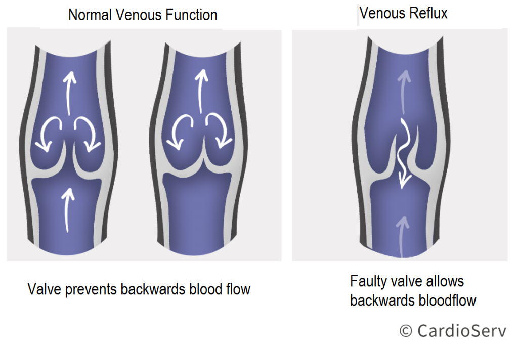
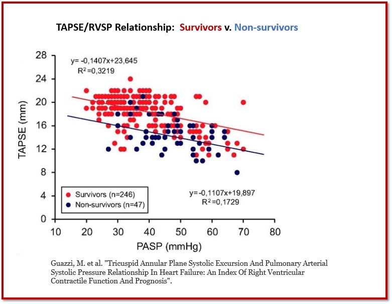
Cerebrovascular Testing
WHAT IS CEREBROVASCULAR TESTING?
Signs & Symptoms
Patient History
Mechanisms
Physical Exam
Cerebrovascular Testing Read More »
WHAT IS CEREBROVASCULAR TESTING?
Signs & Symptoms
Patient History
Mechanisms
Physical Exam
Cerebrovascular Testing Read More »
Now that we have covered all the methods for evaluating mitral regurgitation (MR), let’s put it to use! The ASE released an algorithm as a guide to help determine the severity of chronic MR, using both qualitative, semi-quantitative & quantitative measures.
ASE’s Mitral Regurgitation Algorithm Simplified! Read More »
This week we are going to cover our last of the three quantification methods for evaluating the severity of mitral regurgitation (MR). The past two weeks we discussed PISA and stroke volume methods. If you missed them or would like to refresh, you can find them there:
Volumetric Method: A Third method for evaluating MR severity! Read More »
As technology advances and we are encouraged to implement additional measurements into our routine scanning exams, it’s good to take a step back and refresh on proper techniques! Our goal for this blog is to have a better understanding of proper technique for measuring left ventricle (LV) volumes!
Let’s Talk Left Ventricle Bi-Plane Volume Measurements! Read More »
Last month in July, CardioServ partnered with All About Ultrasound to hold our Mastering Venous Reflux Ultrasound Conference. Our goal at CardioServ is to inspire excellence in imaging. This includes making sure we teach to all different learning styles. We have become more aware, over the past years, of just how differently our clients learn and process information.
Missed our Ultrasound Conference? Watch our video review! Read More »
We started our mitral regurgitation (MR) blog series to provide our readers a clear & easy understanding of the basics to evaluating the mitral valve structure when MR is present. If you need refreshing or missed the blogs, you can find them here:
3 Essential Color Doppler Measurements for MR Read More »
We started our mitral regurgitation (MR) blog series with the intention to provide our readers a clear & easy understanding of the basics to evaluating the mitral valve structure when MR is present. Starting next week, we are now going to move on to the suggested methods to qualifying & quantifying the severity of MR according to the recent ASE guidelines that were published this year, Recommendations for Noninvasive Evaluation of Native Valvular Regurgitation.
7 Factors that Influence Color Doppler Appearance Read More »
Let’s talk about some basic hemodynamics that take affect when evaluating mitral regurgitation (MR). We previously broke down the step by step process the heart encounters when dealing with acute & chronic MR. If you’ve missed or need refreshed on our previous blogs, you can find them here:
FYI: Temporal Variation & Blood Pressure Matters for MR! Read More »
Last week we discussed clear & easy tips on how to evaluate different abnormalities of the mitral valve (MV) for primary MR. We also explained the Carpentier’s Classification concept and how it’s applied to various etiologies of the MV. If you need to refresh or missed our past blogs, you can find them here:
Secondary MR: Evaluating Mitral Valve Tethering Read More »
The ASE recently released their new 2017 valvular regurgitation updated guidelines back in April. The paper details the difference between primary and secondary mitral regurgitation (MR) and outlines proper evaluation methods for the most common pathologies seen. The past couple of weeks, we have discussed how to differentiate between primary vs. organic and acute vs. chronic. If you need to refresh or missed our blog, you can read them here:
Essential Steps to Evaluating MR Etiologies Read More »