
Last Updated on January 30, 2024 by Hannes van der Merwe
We started our mitral regurgitation (MR) blog series to provide our readers a clear & easy understanding of the basics to evaluating the mitral valve structure when MR is present. If you need refreshing or missed the blogs, you can find them here:
- Mitral Valve Anatomy: Name 5 Components!
- Finally… Mitral Valve Orientation Explained!
- Smart Strategies for Determining MR Mechanisms
- Ultimate Guide to Acute vs. Chronic MR
- Essential Steps to Evaluating MR Etiologies
- Secondary MR: Evaluating Mitral Valve Tethering
- FYI: Temporal Variation & Blood Pressure Matters for MR!
- 7 Factors that Influence Color Doppler Appearance
Starting this week, we are now going to move into the suggested methods for qualifying & quantifying the severity of MR according to the recent ASE guidelines that were published this year, Recommendations for Noninvasive Evaluation of Native Valvular Regurgitation. Our goal for this blog is to provide qualitative & semi-quantitative methods using color Doppler to evaluate MR.
COLOR DOPPLER
Color flow Doppler is the primary method to evaluating the presence of regurgitation. It provides us information on:
- Location of regurgitation jet
- Size of jet
- Spatial orientation of jet area (width & length)
- Flow convergence into regurgitant orifice
We tend to rely on color Doppler to determine severity of a regurgitant jet as our primary source. This is not the best practice for quantifying the severity of MR. Remember, color Doppler is a primary method to evaluate the presence of regurgitation, not the severity of MR.
COLOR DOPPLER METHODS
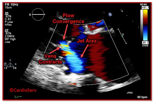
There are 3 components of a color Doppler jet that make up the appearance we see on the screen. When we use color Doppler for evaluation of regurgitation, it is vital we visualize all 3 parts in order to properly quantify the severity.
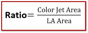
- Color Jet Area
- Vena Contracta (VC)
- Flow Convergence
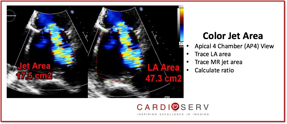
COLOR JET AREA
- Easy to measure
- Good for excluding presence of MR
- Not reliable for grading severity of MR
- Qualitative method
- Limitations: inconsistent between follow-up studies & dependent on hemodynamics

Limitations:
- Inconsistent between follow-up exams
- Dependent upon hemodynamics
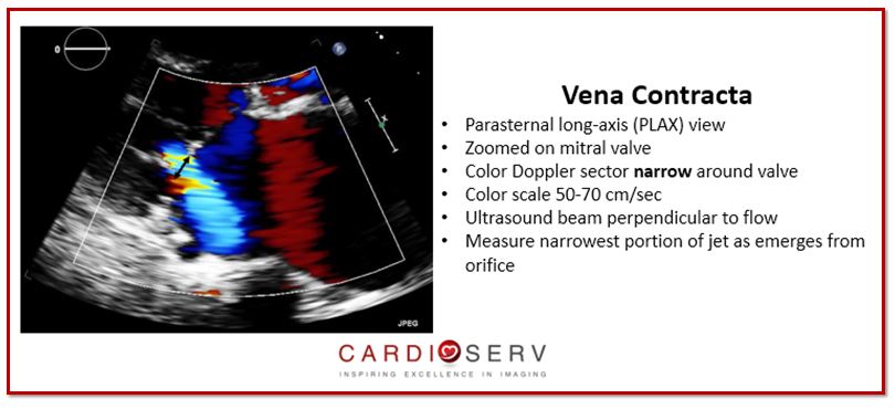
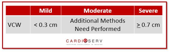
VENA CONTRACTA
- Narrowest portion of jet as emerges from orifice
- Good marker to distinguish mild from severe MR
- Semi-quantitative measure
- Independent of flow rate & driving pressures of a fixed orifice
Limitations:
- Dependent upon orifice geometry shape
- Multiple jets = underestimation
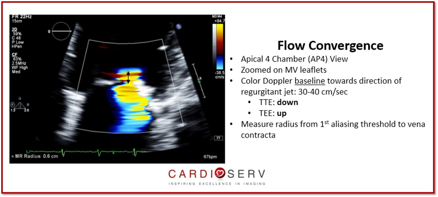
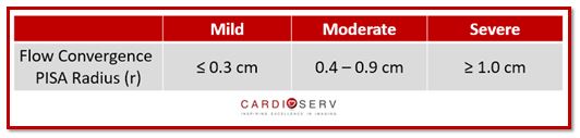
FLOW CONVERGENCE
- Located proximal to regurgitant orifice
- Qualitative measure
- Provides information on location of lesion causing MR & magnitude of regurgitant flow
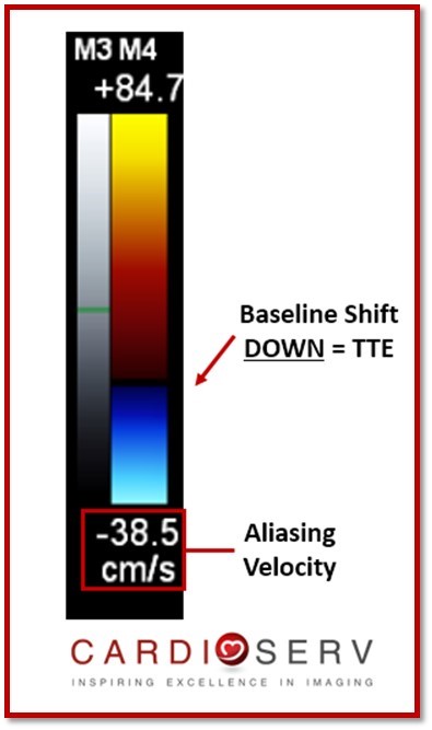
- Radius (r) is the diameter length from point of color aliasing to vena contracta
- Ideal for central regurgitation jets (circular orifices)
- First hemispheric aliasing shell
- Appear as ‘tear-drop’ or ‘golf-tee’
Limitations:
- Secondary MR orifice crescent shape causes underestimation
- Error in radius measurement causes major miscalculation to determine EROA (radius is squared)
- Calculated from single-frame image
- If regurgitation is NOT holosystolic causes overestimation
SUMMARY
This week we reviewed suggested methods for evaluating the presence of MR with color Doppler. We encourage you to incorporate the following into your scanning protocol when MR is detected:
- Color Jet Area
- Vena Contracta
- Flow Convergence
Keep an eye out for upcoming blogs over methods to quantify the severity of MR! Our goal at CardioServ is to provide you with an easy guides to help incorporate positive change & improvement. We look forward to hearing feedback and comments from our readers!

Andrea Fields MHA, RDCS
Stay Connected: LinkedIn, Facebook, Twitter, Instagram
References:
Thomas, J. D., MD, & Liu, C., MD. (1990). Quantification of Jet Flow by Momentum Analysis. Circulation ,81, 1st ser., 247-259. Retrieved July 19, 2017, from http://circ.ahajournals.org/content/81/1/247.long
Zoghbi, W. A., MD, FASE, & Adams, D., RCS, RDCS, FASE. (2017). Recommendations for Noninvasive Evaluation of Native Valvular Regurgitation. JASE, 30, 4th ser., 1-69. Retrieved June 12, 2017.


