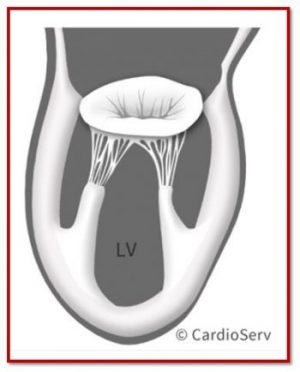
Mitral Valve Anatomy: Name 5 Components!
Last week we wrapped up our right heart blog series. Be sure to keep an eye out for our Right Heart E-Book that will be available soon!
Mitral Valve Anatomy: Name 5 Components! Read More »
Last week we wrapped up our right heart blog series. Be sure to keep an eye out for our Right Heart E-Book that will be available soon!
Mitral Valve Anatomy: Name 5 Components! Read More »
Last week we discussed 3 measures to use with color Doppler for evaluating the presence of mitral regurgitation (MR). If you missed last week’s blog, or would like to refresh, you can find them here:
3 Doppler Techniques for Evaluation of MR! Read More »
Mitral regurgitation (MR) can be quantified several ways in echocardiography, and one of the most practical, reproducible approaches is the Stroke Volume (SV) Method.
3 Steps to Quantifying Mitral Regurgitation via Stroke Volume Method! Read More »
Learn the 3 variables used in quantifying the severity of mitral regurgitation (MR) and take a closer look at the first method for obtaining them — the PISA method.
A Complete Guide to Performing MR PISA! Read More »
Last week, we covered the basic anatomy of the mitral apparatus. We reviewed in detail, the various structures and their function. As we continue with our mitral regurgitation blog series, we want to touch next on, specific imaging windows to evaluate the mitral valve. Remember, when we examine the valve, we need to make sure we include all structures of the apparatus.
Finally… Mitral Valve Orientation in Echo Explained! Read More »