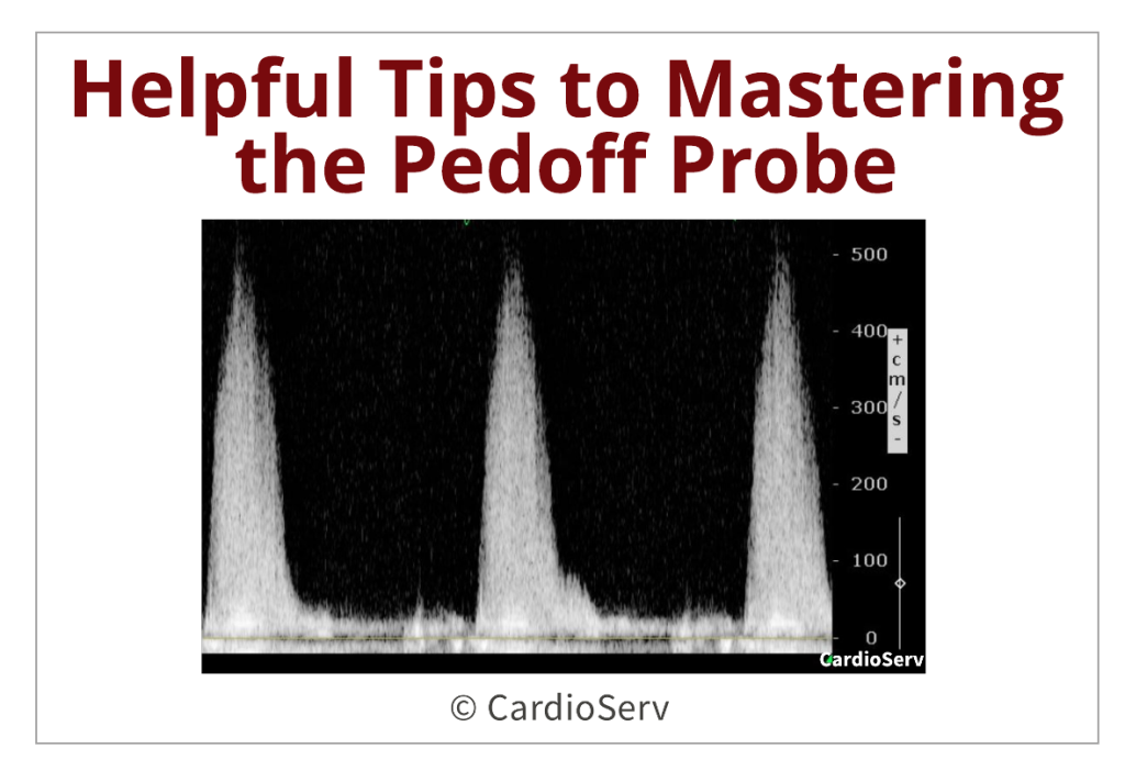
Helpful Tips to Mastering the Pedoff Probe!
Before harmonic imaging, our imaging transducers had major limitations when it came to obtaining high-velocity gradients with continuous-waved (CW) Doppler. Two of the main restrictions included the ultrasound frequency (no ability to drop down to a lower frequency) and the large footprint of the transducer.
Helpful Tips to Mastering the Pedoff Probe! Read More »