
Last Updated on January 30, 2024 by Hannes van der Merwe
We started our mitral regurgitation (MR) blog series with the intention to provide our readers a clear & easy understanding of the basics to evaluating the mitral valve structure when MR is present. Starting next week, we are now going to move on to the suggested methods to qualifying & quantifying the severity of MR according to the recent ASE guidelines that were published this year, Recommendations for Noninvasive Evaluation of Native Valvular Regurgitation.
This week we will lay the foundation for a better understanding of the role of color Doppler with regurgitant jets. Our goal for this blog is to:
- Understanding the role momentum plays in color Doppler jet size
- Optimizing Color Doppler
COLOR DOPPLER
Color flow Doppler is the primary method to evaluating the presence of regurgitation. The obvious benefits of color Doppler include the identification of:
- Location and size of regurgitation jet origin
- Spatial size of jet area (in relation to the receiving chamber)
- Flow convergence into regurgitant orifice
We tend to rely on color Doppler to determine severity of a regurgitant jet as our primary source. This is not the best practice for quantifying the severity of MR. Remember, color Doppler is a primary method to evaluate the presence of regurgitation, not the severity of MR.
MOMENTUM AND JET SIZE
Regurgitant jets are composed of the transfer of mass, energy and momentum into the receiving chamber. The conservation of these principles control the behavior of the jet.
- Conservation of mass (continuity equation)
- Conservation of energy (Bernoulli equation)
- Conservation of momentum
We easily forget that momentum plays a major role in the size of a color Doppler regurgitant jet. Within cardiac ultrasound we are familiar with the continuity equation (mass) and the Bernoulli equation (energy). These two formula’s alone though, cannot predict the size of a color jet – momentum is a determining factor to the size of a color jet.
The geek take away is:
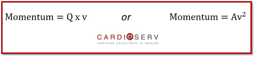
Q = Flow Rate ; v = Velocity ; A = Orifice Area
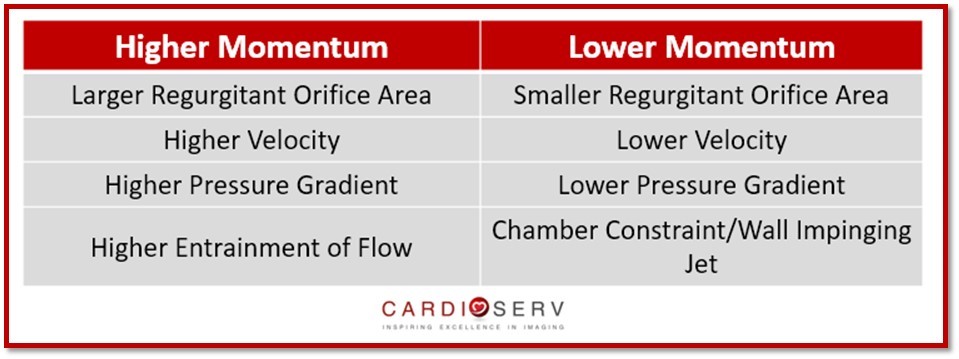
The real world take away from this is understanding that higher momentum will increase jet area and lower momentum will decrease jet area.
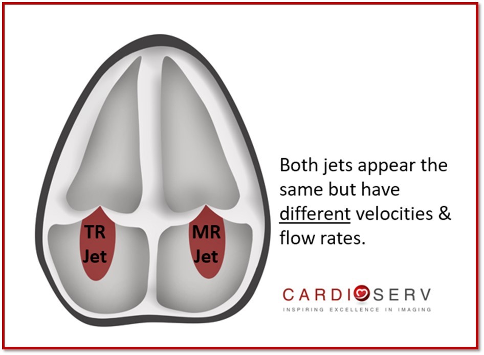
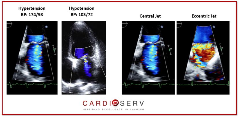
This is why the size of a color jet are the same with a 5m/s MR jet with a flow rate of 100mL as a 2.5m/s TR jet with a flow rate of 200mL/sec. They have the same jet area even though they have different flow rates and velocities. This is a prime example of why we cannot diagnose regurgitation severity based off of color alone.
Now with a little understanding of momentum, it will help us remember that the size and appearance of the color jet area is affected by:
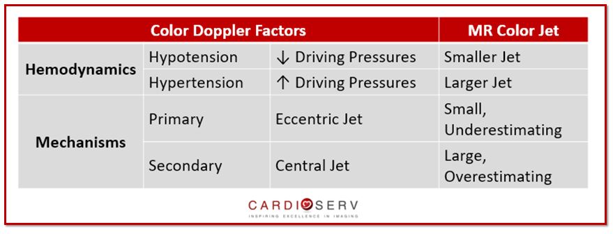
- Hemodynamics: driving pressures (blood pressure)
- Mechanisms: primary vs secondary
If you missed our prior blogs covering these topics, you can find them here:
- FYI: Temporal Variation & Blood Pressure Matters for MR!
- Smart Strategies for Determining MR Mechanisms!
OPTIMIZING COLOR DOPPLER
These two basic settings will help optimize the presence of regurgitation:
- Nyquist limit (aliasing velocity) set between 50-70 cm/sec
- High color gain (Turn your gains up high then lower the just enough to eliminate ‘noise’ from non-moving objects)
7 FACTORS THAT INFLUENCE COLOR DOPPLER APPEARANCE
- Valve Structure: color jet will flow opposite direction of the abnormal leaflet
- Blood Pressure (momentum): larger color jet with increased driving pressures
- Color Scale: decrease color scale will emphasize lower velocities
- PRF: increase PRF = decrease jet area size
- Color Doppler Gains: decrease gains = diminish sensitivity causing large jets to appear smaller
- Attenuation: structures in the far field or metal objects will cause a decrease in jet size appearance
- Frequency: TEE has higher frequencies which causes lower velocity jets to appear larger
Keep an eye out for upcoming blogs over methods to quantify the severity of MR! Our goal at CardioServ is to provide you with an easy guides to help incorporate positive change & improvement within the imaging labs. We look forward to hearing feedback and comments from our readers!

Andrea Fields MHA, RDCS
Stay Connected: LinkedIn, Facebook, Twitter, Instagram
References:
Thomas, J. D., MD, & Liu, C., MD. (1990). Quantification of Jet Flow by Momentum Analysis. Circulation ,81, 1st ser., 247-259. Retrieved July 19, 2017, from http://circ.ahajournals.org/content/81/1/247.long
Zoghbi, W. A., MD, FASE, & Adams, D., RCS, RDCS, FASE. (2017). Recommendations for Noninvasive Evaluation of Native Valvular Regurgitation. JASE, 30, 4th ser., 1-69. Retrieved June 12, 2017


