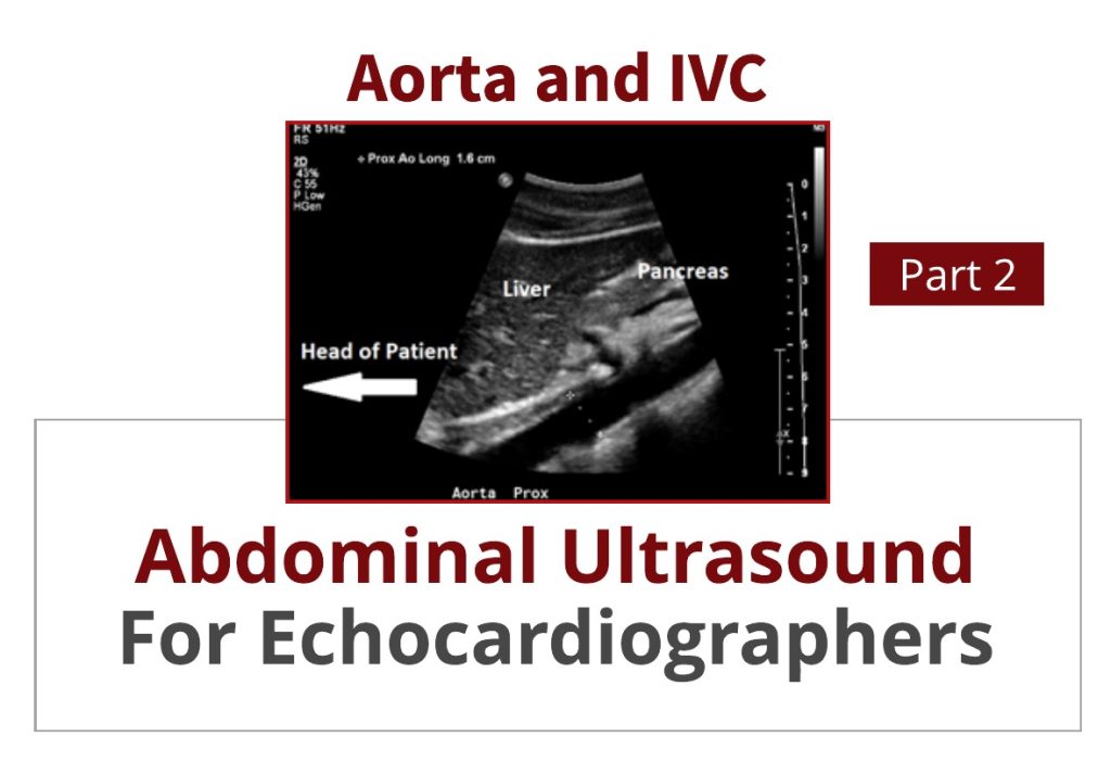
Abdominal Ultrasound for Echocardiographers: Aorta and IVC
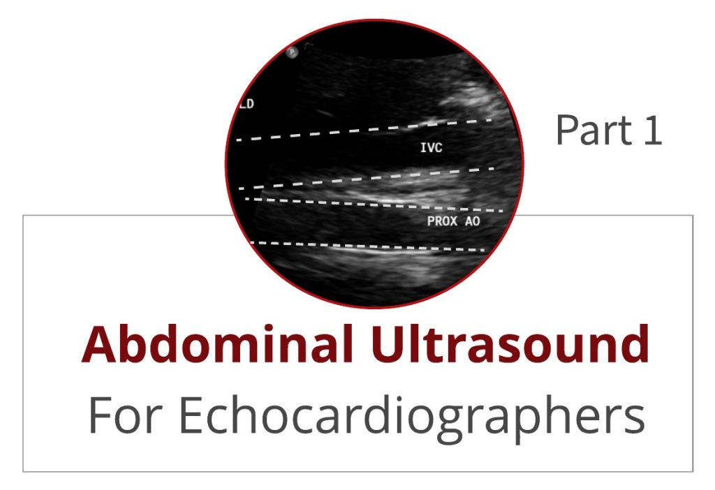
In an early blog, Abdominal Ultrasound for Echocardiographers: Part 1, we reviewed some basic tips for echocardiographers scanning the abdomen. We reviewed artifacts, image orientation and patient positioning.
Abdominal Ultrasound for Echocardiographers: Aorta and IVC Read More »