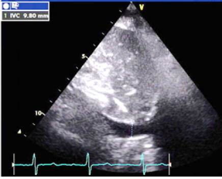
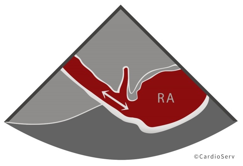
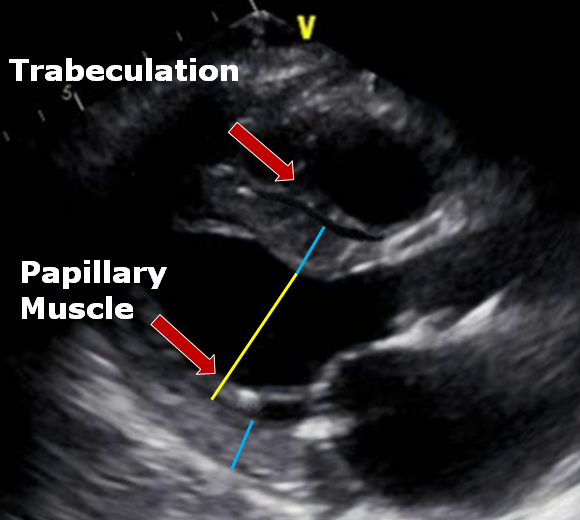
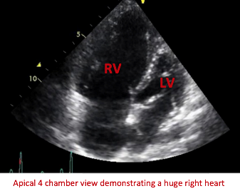
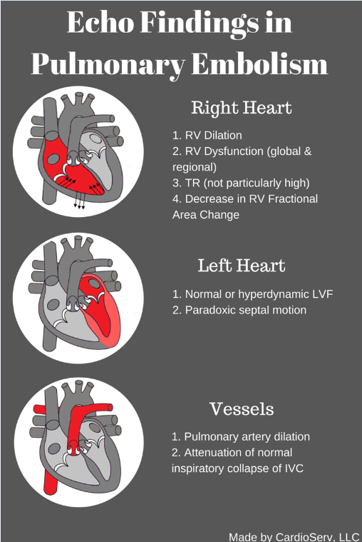
Name That Pathology
Modality: Adult Echo TEE
Review the following images and see if you know what pathology is present! TEE images of LA and RA Color Doppler over the atrial septum demonstrating PFO.
Name That Pathology Read More »
Modality: Adult Echo TEE
Review the following images and see if you know what pathology is present! TEE images of LA and RA Color Doppler over the atrial septum demonstrating PFO.
Name That Pathology Read More »
As diagnostic imaging professionals, we often perform echocardiograms on patients with hypertension to monitor the thickness, strength and wall motion of the heart. Last week we launched our two-part blog on left ventricular hypertrophy (LVH). In part one we explained the pathophysiology behind the various categories of LVH along with the echocardiographic findings. We discussed how LVM and RWT plays a role in identifying the categories of LVH. This week we will answer the question: What is LVM and RWT and how do we obtain these values?
Understanding LVH Part 2: How to Measure LV Mass and Diagnose LVH Read More »
Last month was designated American Heart Month to raise awareness of cardiovascular disease, the leading fatality of Americans. AHA encourages our patients to take control over understanding their risk factors of heart disease including knowing their numbers related to blood pressure. Untreated hypertension can have a long-term effect on the heart causing hypertensive heart disease, which can include coronary heart disease (CAD), left ventricular hypertrophy (LVH) and hypertrophic cardiomyopathy (HCM).
Understanding LVH Part 1: Concentric, Eccentric and Concentric Remodeling Read More »
HOW COULD I IMPROVE THIS IMAGE?
Modality: Transthoracic Echocardiography
View: Subcostal long axis IVC
ANSWER:
The image could be optimized to elongate the IVC to ensure the correct diameter is measured.
Inspiring Excellence Series Read More »
Last week, we shared our new year resolutions which including writing more and inspiring more. So I am jumping right in by writing a blog on the correct method to estimate right atrial pressures (RAP) when calculating your RVSP during an echo. The American Society of Echocardiography has outlined the correct use of the Collapsibility Index to estimate RAP yet the majority of the echocardiograms that we review on a daily basis still have an RAP of 10 entered for every echo. After speaking with sonographers, most admit it’s either a bad habit or it is just what they were taught to do.
8 Things to Know About Estimation of RAP via Echocardiography Read More »
ECHO AND VASCULAR LABS – ARE YOU READY FOR THE NEW SONOGRAPHER CREDENTIALING REQUIREMENTS?
As you may be aware in January 2017 the latest changes to the Echo and Vascular accreditation standards as set forth by the Intersocietal Accreditation Commission become effective. We have discussed the changes to the Quality Improvement Program in recent blogs. In this blog we will cover the changes to the sonographer credentialing and training requirements.
IAC’s New Sonographer Credentialing Requirement Read More »
In our last post we reviewed the correct techniques for the linear measurement of the LV in the parasternal long axis view. This week we will discuss the aorta and review the morphology and the correct aortic measurement techniques to perform during an echocardiogram.
How to Master Aortic Measurements with These 5 Techniques Read More »
Last year the American Society of Echocardiography (ASE) released updated guidelines for cardiac chamber quantification by echocardiography. The ASE teamed up with the European Association of Cardiovascular Imaging to update the previous guidelines due to the rapid technological developments of the past decade and the changes in echocardiographic practice brought about by these developments.
6 Pitfalls of Accurate LV Measurements Read More »
For those following our blog series you should be getting the hang of streamlining your QI Program by now. This week we will tackle the dreaded clinical correlation! Wait… before you cringe and close out this blog let me share some good news! IAC recently changed the correlation requirements for many of the modalities.
The 4 Most Effective Tactics for Gathering Clinical Correlation Read More »
An 85 year old patient presents to the ER with worsening shortness of breath and a history of known pulmonary hypertension. Her chest x-ray reveals massive cardiomegaly. An echo is ordered to assess the status of her pulmonary hypertension. We will present this echo to illustrate the correct techniques when performing an echocardiogram with the indication of pulmonary hypertension. Learn about the LV Eccentricity index and the latest recommendations for calculating correct RA pressure through evaluation of the IVC.
Pulmonary Hypertension Case Study Presentation Read More »