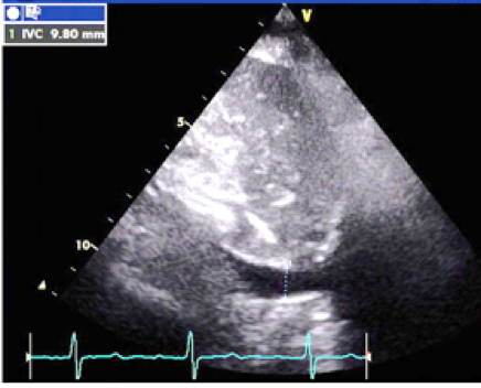
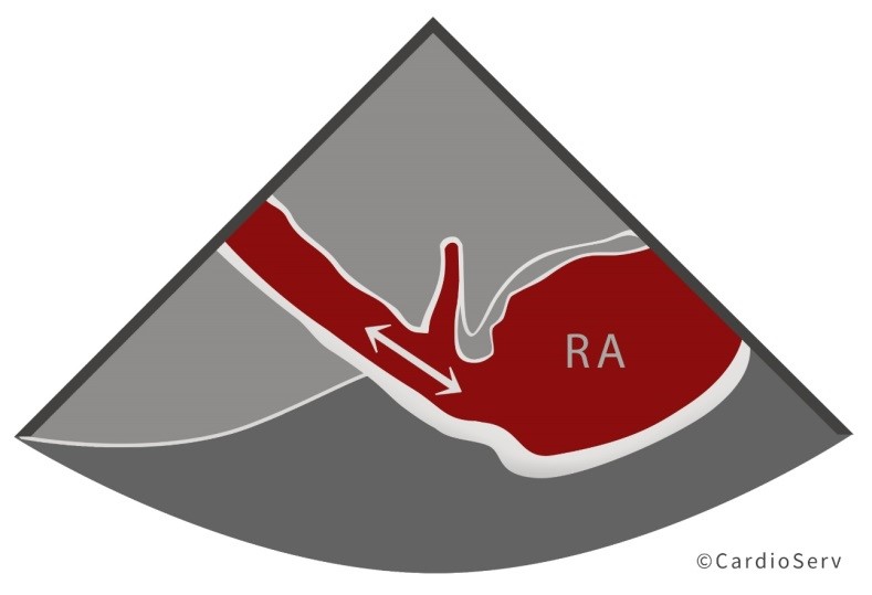
Written by Judith Buckland, MBA, RDCS, FASE
After our last post on 'how to estimate the right atrial pressure', we received questions from our readers. What do we do if we cannot assess the IVC? What if the patient is on a vent? This week we will answer those questions.
Review prior article for information on:
Why estimate...
Written by Yvonne Prince ACS, RDCS, RVT, RDMS, FASE
In an early blog, Abdominal Ultrasound for Echocardiographers: Part 1, we reviewed some basic tips for echocardiographers scanning the abdomen. We reviewed artifacts, image orientation and patient positioning. This week we will provide you with 6 steps to successfully identify the aorta and...
Written by Yvonne Prince ACS, RDCS, RVT, RDMS, FASE
How often have you found yourself “in over your head” in the abdomen when trying to image the IVC and abdominal aorta? Is imaging the abdominal aorta part of your echo protocol? It is not uncommon for a patient to receive an abdominal ultrasound because the echo findings mentioned the...
Written by Andrea Fields MHA, RDCS
Us clinicians commonly ask the same popular question in our field, "What is the cut-off ranges for pulmonary hypertension?!" We want to help better understand the basics of determining the presence of pulmonary hypertension with echocardiography.
First let's start with one important key item to...
Written by Andrea Fields MHA, RDCS
In Hepatic Veins 101, we discussed the structure, function and waveforms of the hepatic veins, in regards to the cardiac cycle. This blog will cover how the hepatic veins play a role with the right heart. We tend to forget about the importance and useful information the hepatic veins provide us....