Last Updated on March 16, 2026 by Don Gerig, RDCS
Mitral regurgitation (MR) can be quantified several ways in echocardiography, and one of the most practical, reproducible approaches is the Stroke Volume (SV) Method.
While the PISA method often takes center stage, the stroke volume approach provides an excellent alternative, especially when color Doppler or aliasing limits PISA accuracy. This guide walks you through how to calculate regurgitant volume (RVol) and effective regurgitant orifice area (EROA) using the stroke volume method, step-by-step and aligned with ASE recommendations.
Our goal, as always, is to make complex echo concepts simple and practical. By the end of this post, you’ll not only understand the stroke volume method for quantifying MR, but you’ll also see how it fits into your daily workflow and be ready to implement it confidently in your echo lab.
The Concept Behind the Stroke Volume Method
This method is based off of the regurgitant volume (RVol) calculation: Difference between the inflow and outflow stroke volumes (SV) of the left ventricle (LV).
Concept based on: if there is no regurgitation present, the stroke volume at both sites are equal.
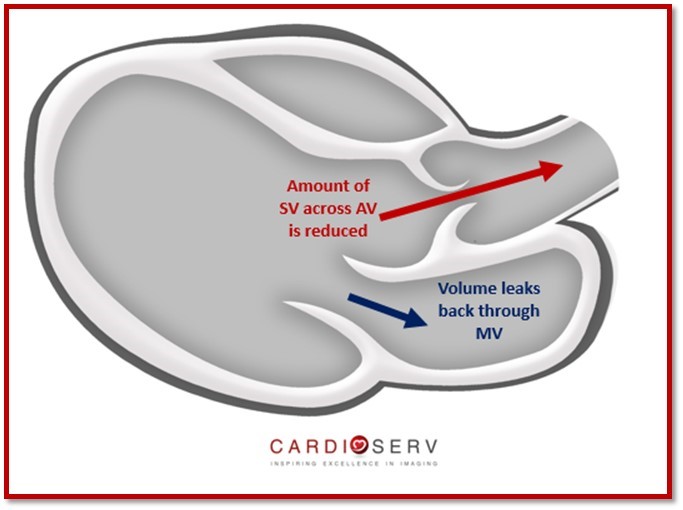
When MR is present, part of the left ventricular stroke volume leaks backward through the mitral valve instead of flowing forward through the LVOT. The difference between these two volumes represents the regurgitant volume (RVol).
The Stroke Volume Method Give You:
- Regurgitant Volume (RVol): LV inflow SV − LV outflow SV
- Regurgitant Fraction (RF): RVol ÷ LV inflow SV × 100
- Effective Regurgitant Orifice Area (EROA): RVol ÷ MR VTI
Understanding Stroke Volume
Stroke Volume is the amount of blood (volume) pumped by the heart with each beat. In a heart with normally functioning valves, absent of regurgitation, the volume entering the left ventricle across the mitral valve (inflow) is equal to the volume exiting the left ventricle across the aortic valve (outflow). Meaning, the SV across the aortic valve equals the SV across the mitral valve.
Stroke Volume in The Presence of Mitral Regurgitation
In cases of mitral regurgitation, however, the forward SV (through the aortic valve) is reduced because part of the LV output regurgitates back through the mitral valve.
This difference in volume is how the regurgitant volume is calculated. Lets review how to calculate the inflow and outflow stroke volumes and learn how to use this information to calculate the regurgitant volume.
How Is Stroke Volume Calculated?
The two valves we will be focusing on to determine the SV method for MR are the Mitral Valve (inflow) and the Aortic Valve (outflow).
You can calculate the SV of an orifice by obtaining two measures:
- Cross-sectional Area (CSA)
- Inflow VTI at the Annulus

It’s important to know that we must obtain the measurement for both values at the same location. We calculate the SV for each valve during their normal flow period:
- Mitral Valve: Diastole
- Aortic Valve: Systole
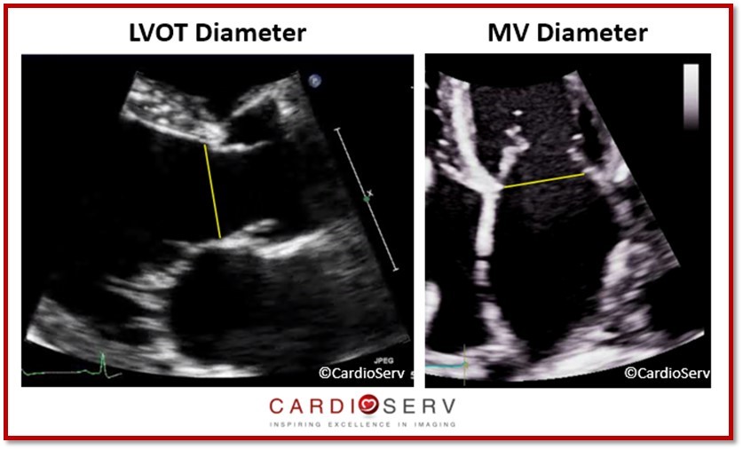
Cross Sectional Area (CSA)
This method assumes the annulus is circular. We can calculate the CSA of an annulus by obtaining the diameter at the location.
- Mitral Valve: Early-Diastole & Inner-edge to Inner-edge
- Aortic Valve (LVOT Diameter): Mid-Systole & Inner-edge to Inner-edge

Velocity-Time Integral (VTI)
For each annulus, we measure the pulsed-wave (PW) Doppler at the same location as where the diameter measurement was taken.
- Mitral Valve (Diastole)– above the baseline
- Aortic Valve [LVOT] (Systole)– below the baseline
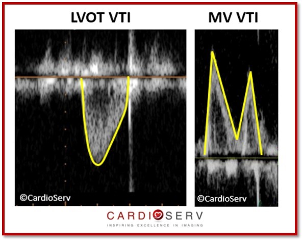
Key Tip: Place the sample volume at the SAME location as diameter was taken. This is vital for the MV because we are use to placing the sample volume at the leaflet tips to evaluate diastolic function–however: for determining MV SV, we must place the sample volume gate at the MV annulus!
Stroke Volume Calculation
Once you have CSA and VTI for each valve:
- Mitral SV = MV CSA × MV VTI
- Aortic SV = LVOT CSA × LVOT VTI
Putting It All Together: Step-by-Step
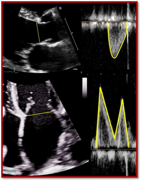
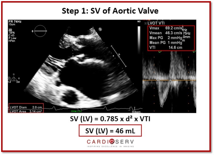
Step 1: Measure Aortic (LVOT) Outflow Stroke Volume
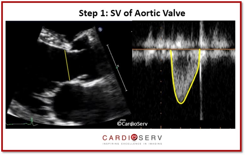
- Parasternal Long Axis view (PLAX): LVOT Diameter (mid-systole)
- Apical 5 chamber or 3 chamber: obtain LVOT PW Doppler
- Record LVOT VTI
Equation: SV (aortic) = π × (LVOT diameter ÷ 2)² × LVOT VTI
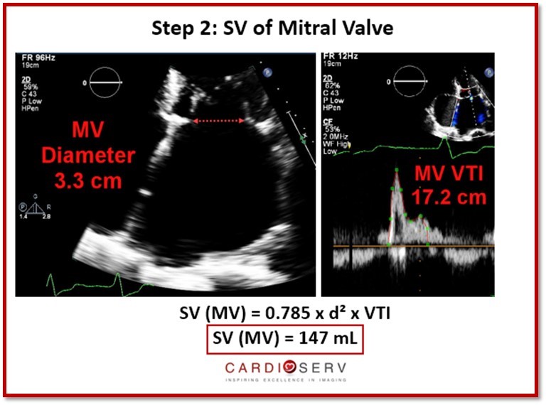
Step 2: Measure Mitral Valve Inflow Stroke Volume
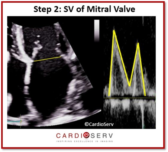
- Apical 4-chamber: measure MV annulus diameter (early diastole)
- Place PW Doppler at the annulus (not leaflet tips)
- Record MV inflow VTI
Equation: SV (mitral) = π × (MV diameter ÷ 2)² × MV VTI
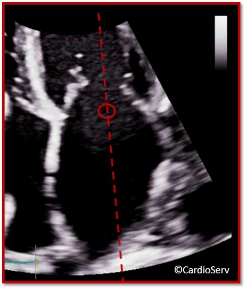
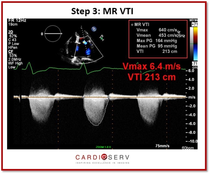
Step 3: Measure MR Jet Velocity (CW Doppler)
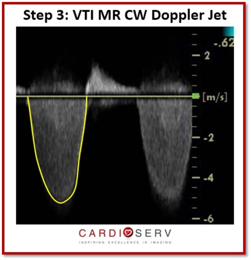
- Zoom the mitral valve and left atrium
- Activate color Doppler and trace the MR jet
- Use CW Doppler to measure MR VTI
Then calculate:
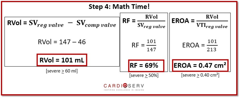
- Regurgitant Volume (RVol) = MV SV − LVOT SV
- Regurgitant Fraction (RF) = RVol ÷ MV SV × 100
- EROA = RVol ÷ MR VTI
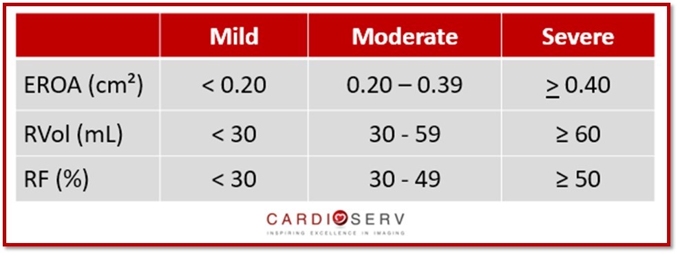
Interpretation of Quantitative MR Severity Using SV (ASE Criteria)
Common Pitfalls and Limitations
- Incorrect annular measurement (use the same site for diameter and Doppler)
- Poor Doppler alignment: underestimates VTI
- Noncircular annulus (especially mitral): introduces geometric error
- Arrhythmias or beat-to-beat variability: average multiple cycles
Case Example
Now that we’ve discussed how to perform this method of quantification, let’s break down a case example!
Final Thoughts and Next Steps
The Stroke Volume Method is one of the most reliable quantitative tools for assessing mitral regurgitation, especially when PISA isn’t feasible. By measuring flow at both the mitral and aortic valves, you can determine true regurgitant volume and severity using data already obtainable in most echo labs.
When performed carefully and consistently, this approach enhances diagnostic confidence, improves reproducibility, and supports IAC and ASE-compliant quantification standards.
We appreciate all of our readers and love to hear feedback! Be sure to check out our other articles!
Take the Guesswork Out of Mitral Valve Imaging
Mitral Valve Imaging in Echo – Visual Guide
Sharpen your MR assessment with this clear, image-rich guide to echo views, scallops, and probe positioning.
- ✔ Understand scallop anatomy in TTE & TEE with ease
- ✔ Avoid common pitfalls like foreshortened views
- ✔ Dozens of labeled diagrams for quick reference
- ✔ Practical checklists to streamline MR imaging

Stay Connected: LinkedIn, Facebook, Twitter, Instagram
References:
Zoghbi, W. A., MD, FASE, & Adams, D., RCS, RDCS, FASE. (2017). Recommendations for Noninvasive Evaluation of Native Valvular Regurgitation. JASE, 30, 4th ser., 1-69. Retrieved June 12, 2017.


