Last Updated on March 24, 2026 by Don Gerig, RDCS
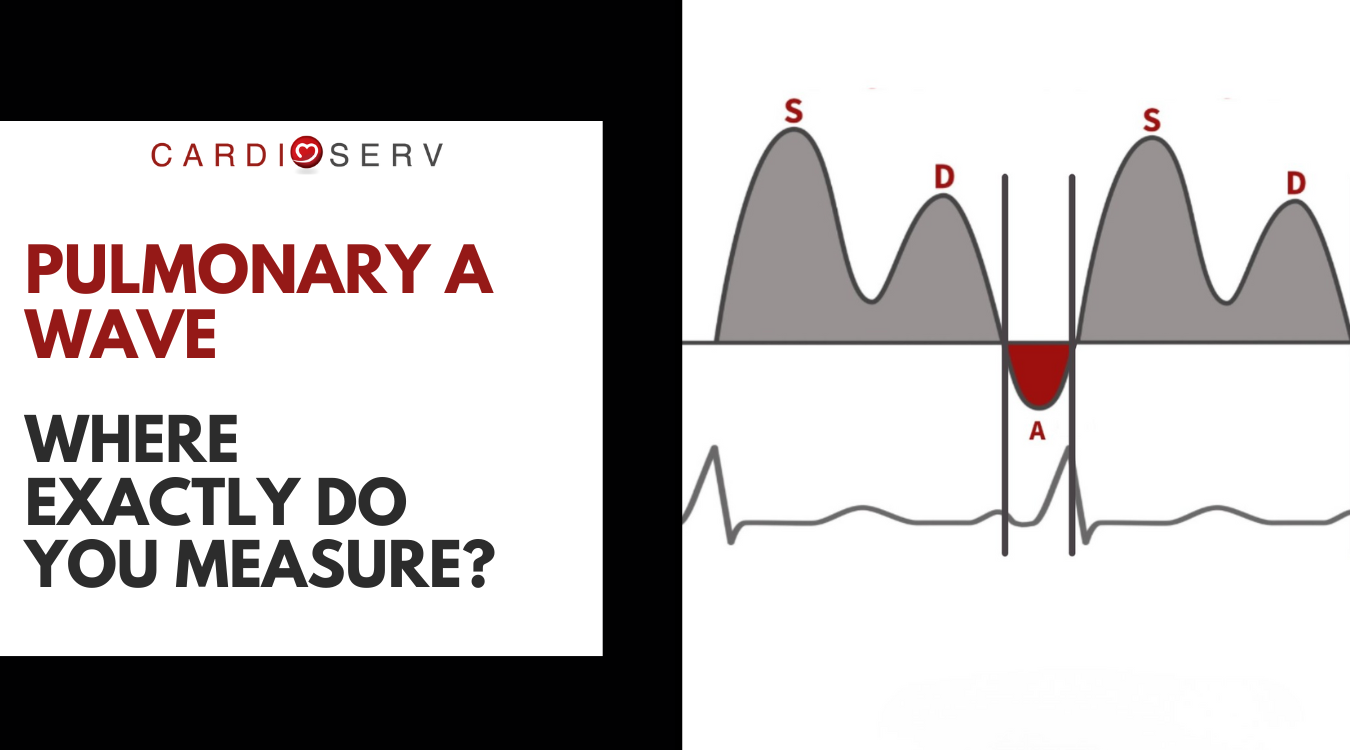
A common questions we’ve received since the rollout of updated diastolic function protocols is about pulmonary venous Doppler — specifically:
“Are the A wave and AR duration measured before the S wave or after the D wave? I’ve seen it shown both ways, and I’m getting conflicting explanations.”
If you’ve asked this yourself, you’re not alone. Pulmonary veins can be challenging to acquire — and even more confusing to interpret when different diagrams appear to show different timing.
Let’s clear this up once and for all.
Start With the Physiology (This Is the Key)
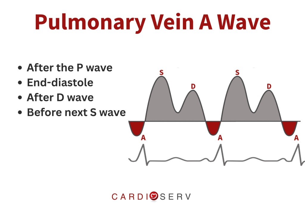
Pulmonary venous atrial reversal (AR) represents left atrial contraction.
That means:
- It occurs after the P wave
- It happens at end-diastole
- It always occurs after the D wave
- It always occurs before the next S wave
This timing does not change.
So if you’re wondering whether AR is “before S” or “after D,” the answer is:
👉 Yes — both are correct, because they describe the same physiologic moment.
Why the Diagrams Look Different (But Aren’t)
ASE examples, IAC images, and educational schematics (including ours) may look different, but they are all illustrating the same atrial contraction event.
What changes is:
- Where the waveform is visually emphasized
- How the cardiac cycle is labeled
- Whether the diagram is conceptual or clinical
What does not change:
- The underlying physiology
- The timing of atrial contraction
- The duration of the AR wave
In other words, this is a display issue, not a measurement rule change.
So What Are You Actually Measuring?
AR Duration =
Onset of atrial reversal → end of atrial reversal
That’s it.
You are not measuring:
- Relative to the S wave
- Relative to the D wave
- Relative to whichever part of the waveform looks “cleanest”
You are measuring:
- Atrial contraction
- Ideally anchored to the ECG P wave
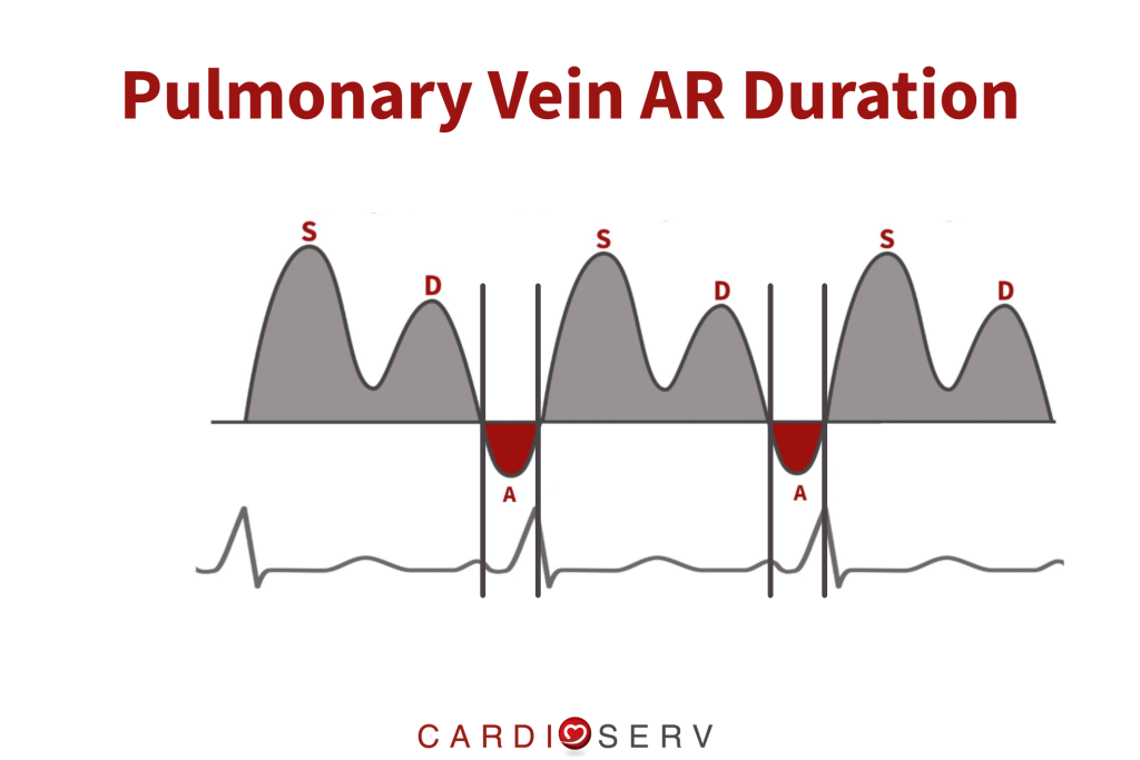
If the AR is correctly identified, its duration will be the same whether your calipers appear:
- after the D wave
or - just before the next S wave
Practical Tips for Sonographers
If pulmonary veins feel intimidating, focus on these fundamentals:
- Use ECG gating whenever possible
- Confirm AR aligns with the P wave
- Measure only the true reversal component
- Ignore baseline noise before or after
- ❌ Don’t overthink where it “sits” visually
If it’s truly atrial contraction, the timing will always make sense.
What Do the Guidelines Say?
- ASE defines pulmonary venous AR as flow reversal during atrial systole
- IAC recognizes AR as an end-diastolic event
- Classic echo literature compares AR duration to mitral A duration, reinforcing that both represent atrial contraction
Across guidelines and research, the message is consistent:
AR is anchored to atrial physiology — not to waveform aesthetics.
Bottom Line
Pulmonary venous AR duration does not change based on whether it’s drawn before S or after D.
It is the same cardiac event, measured the same way, every time.
Once you anchor your measurement to atrial contraction, the confusion disappears — and pulmonary veins suddenly become a lot less scary.
Have a question you’d like us to tackle next?
Send it our way — chances are, you’re not the only one wondering.
💙
Team CardioServ