Last Updated on March 19, 2026 by Don Gerig, RDCS
In a previous article we discussed the algorithm for determining the severity of chronic mitral regurgitation, provided by the ASE! If you missed it, you can find it here!
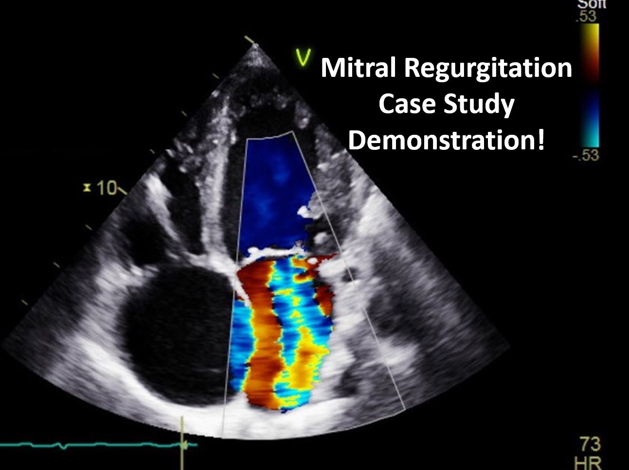
Now let’s apply the ASE algorithm to a real case and walk through how we determine MR severity step-by-step.
Clinical Presentation
A 50 year old male admitted to hospital for shortness of breath and palpitations. EKG showed paced sinus rhythm. BSA is 2.0 m² and blood pressure (BP) is 123/70. The patient has the following history:
- Pacemaker
- Congestive Heart Failure
- Pulmonary Hypertension
- Diabetic
Initial Echo Findings
Parasternal Long Axis
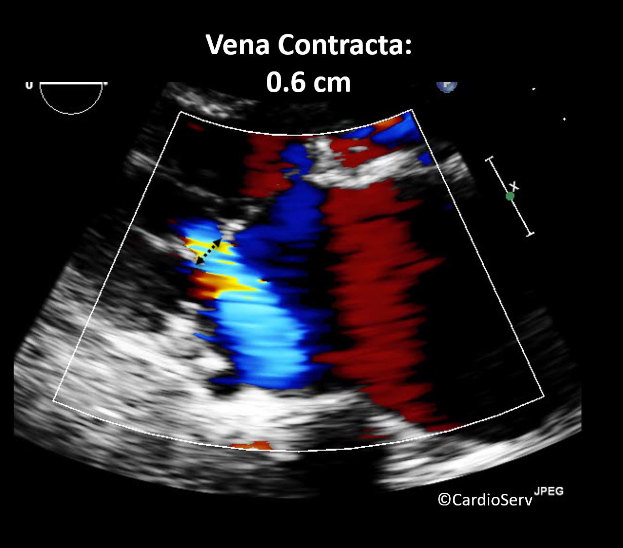
Vena Contracta Width
Apical 4 Chamber (AP4)
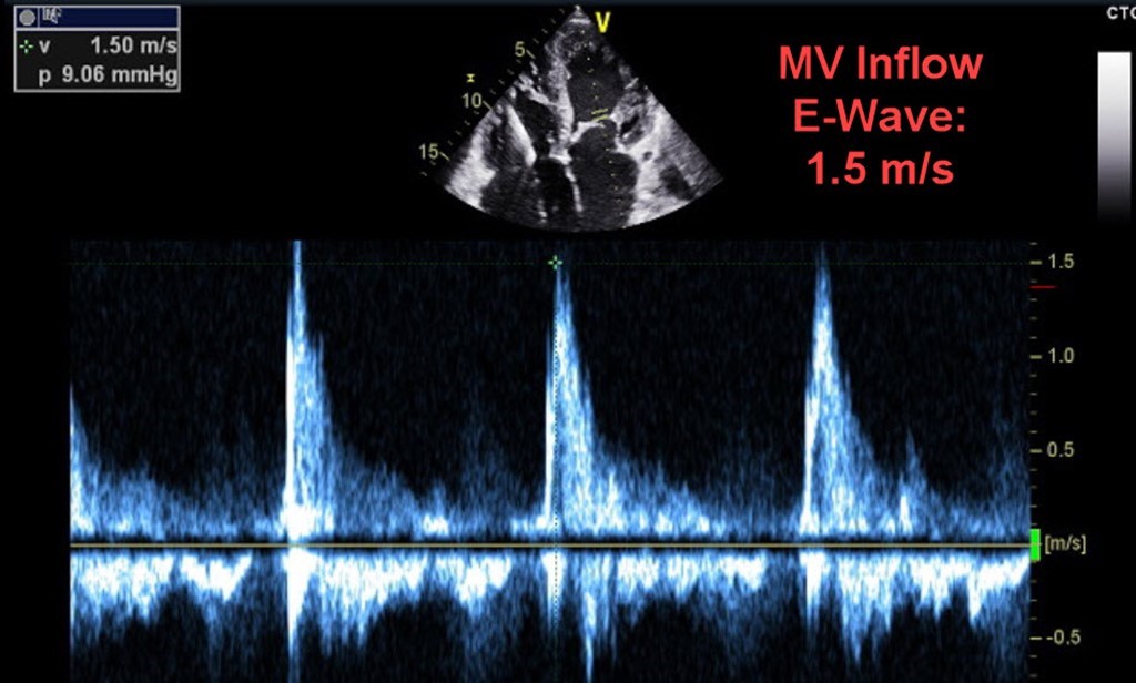
Mitral Valve Inflow
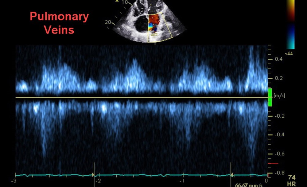
Pulmonary Vein Flow
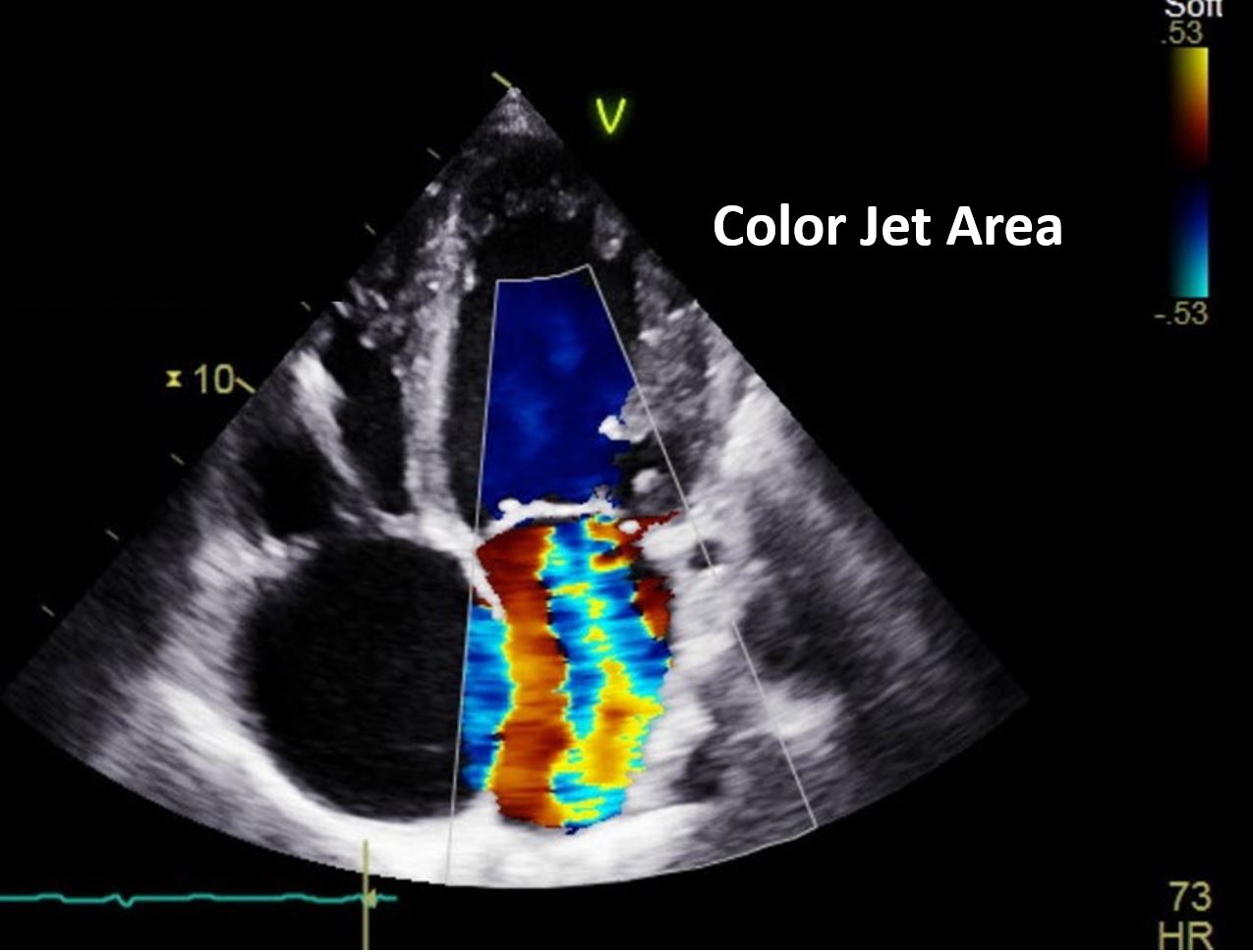
Color Jet Area
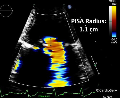
PISA Radius
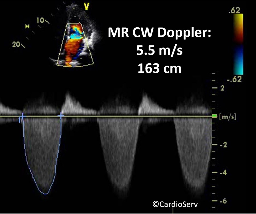
Continuous Wave Doppler
Apical 2 & 3 (AP2 & AP3)
Echo Measurements
- End-Diastolic Volume (EDV) Indexed: 94 mL/m²
- Ejection Fraction (EF): 45%
- LA Volume Indexed: 70 mL/m²
- Vena Contracta Width: 0.6 cm
- MV Inflow E-Wave: 1.5 m/s
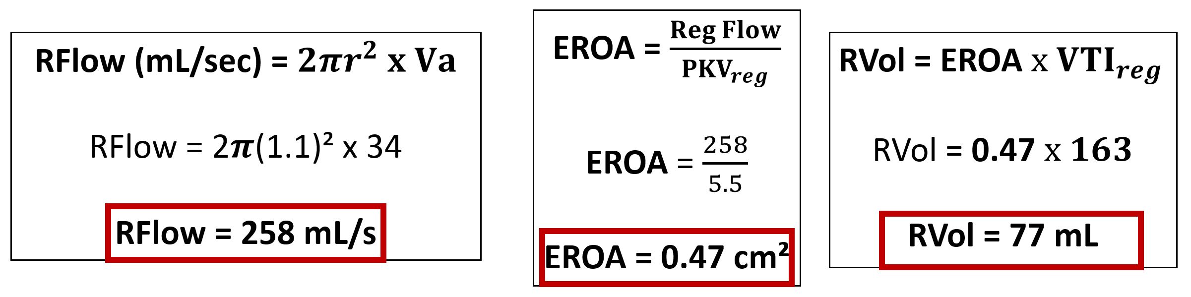
- PISA Radius (Flow Convergence): 1.1 cm
- Aliasing Velocity: 34 cm/sec
- MR VTI: 5.5 m/s & 163 cm
What Do You See?
Before we walk through this case, take a moment to evaluate the findings and ask yourself:
- Based on the images and Doppler, how severe does this MR appear?
- Which parameters stand out most to you?
- What additional measurements would you prioritize?
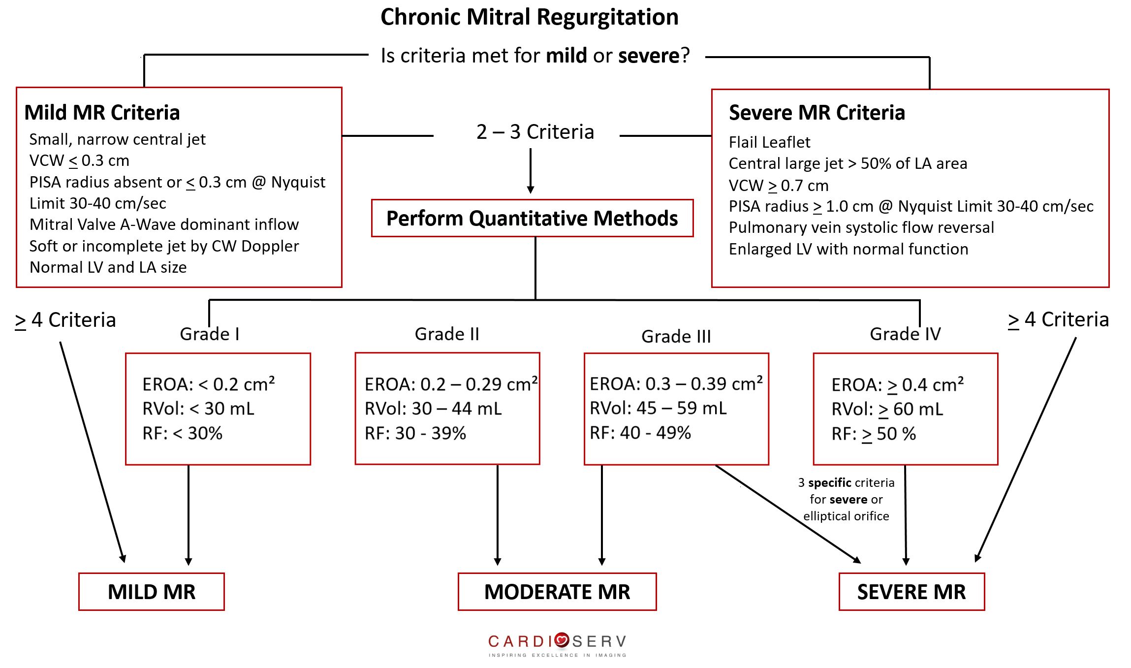
Chronic Mitral Regurgitation Algorithm Chart
For a detailed breakdown of this algorithm chart, read our detailed guide here.
Interpreting the Findings
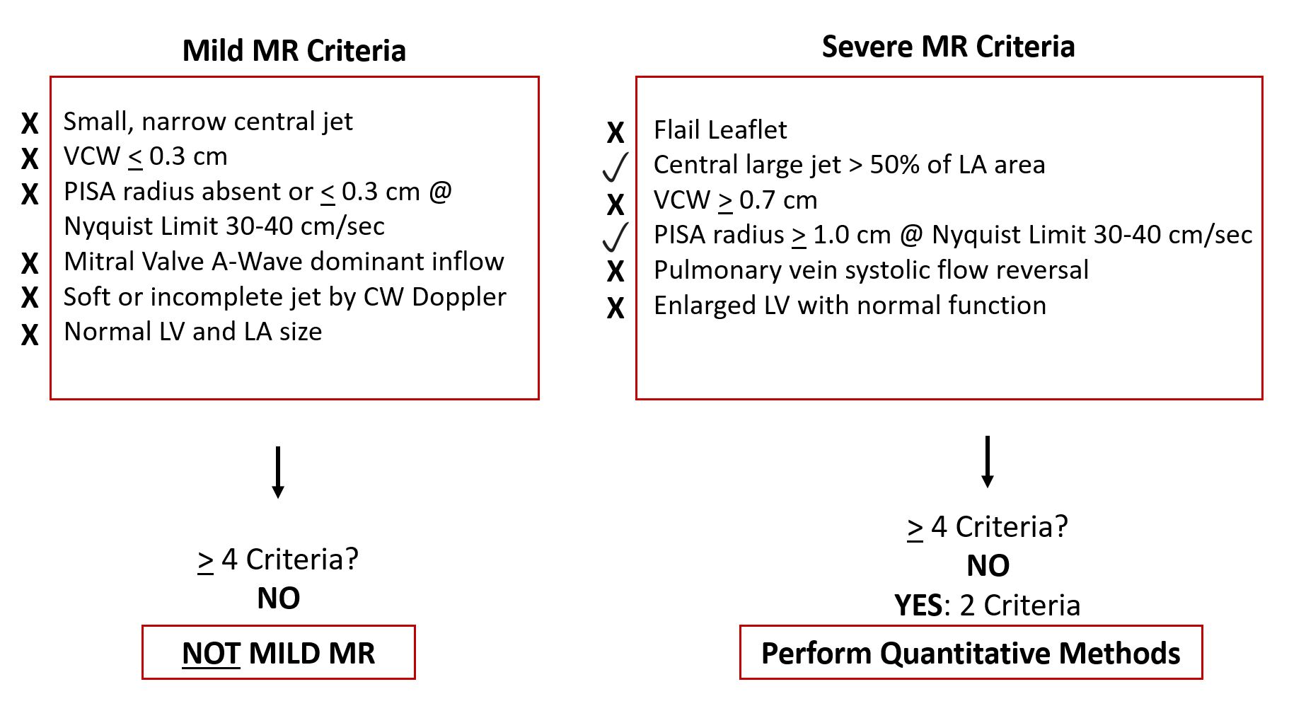
STEP 1: Can We Rule Out Mild or Severe Mitral Regurgitation?
Now let’s apply the ASE algorithm step-by-step to this case:
STEP 2: Perform Quantitative Methods (PISA)
The next step is to calculate PISA which can help determine the severity of mitral regurgitation. For a refresher on calculating PISA check out our guide here.
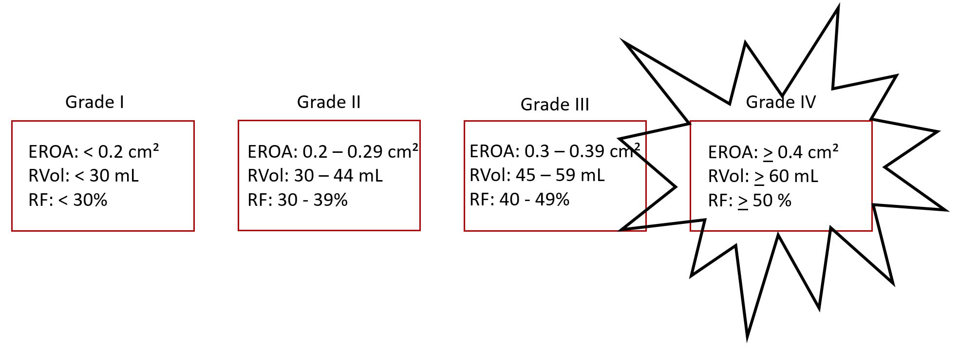
STEP 3: Determine Which Grade The Values Fall Under
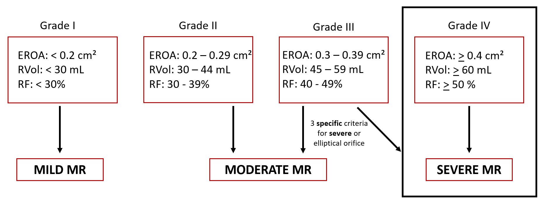
STEP 4: Determine the Severity of Mitral Regurgitation
Final Interpretation
Based on qualitative and quantitative findings, this case is consistent with severe mitral regurgitation.
- Initial findings did not mee 4 of the Mild or Severe criteria, so we moved onto perform quantitative measurements (PISA).
- EROA, RVol and RF were all consistent with Grade IV (Severe) MR
Key Takeaways
- MR severity should never be determined using a single parameter. Always integrate qualitative and quantitative findings.
- Vena contracta and PISA are key but must be interpreted in context. Technical limitations and loading conditions can affect accuracy.
- Pulmonary vein flow reversal strongly supports severe MR. One of the most reliable supportive findings when present.
- PISA-derived values (EROA, regurgitant volume) strengthen severity grading. Especially when findings are discordant.
- Use an integrated, stepwise algorithm approach for consistency. Follow ASE guidance to reduce variability and improve diagnostic confidence.
Go Deeper With Mitral Regurgitation Quantification
Master MR Quantification with Confidence
Advanced Mitral Regurgitation Quantification
A practical, step-by-step course designed to help you understand how key MR measurements work together in real-world echo.
- ✔ Understand Color Doppler, PISA, EROA, RVol, and stroke volume
- ✔ Learn the strengths and limitations of each measurement
- ✔ See how measurements interrelate in a comprehensive approach
- ✔ Apply concepts with clarity in everyday clinical practice

Andrea Fields MHA, RDCS
Stay Connected: LinkedIn, Facebook, Twitter, Instagram
References:
Zoghbi, W. A., MD, FASE, & Adams, D., RCS, RDCS, FASE. (2017). Recommendations for Noninvasive Evaluation of Native Valvular Regurgitation. JASE, 30, 4th ser., 1-69. Retrieved June 12, 2017.


