Last Updated on January 30, 2024 by Hannes van der Merwe
Our next blog series is going to cover the basic material needed to complete a carotid ultrasound study. First, we need to review basic anatomy!
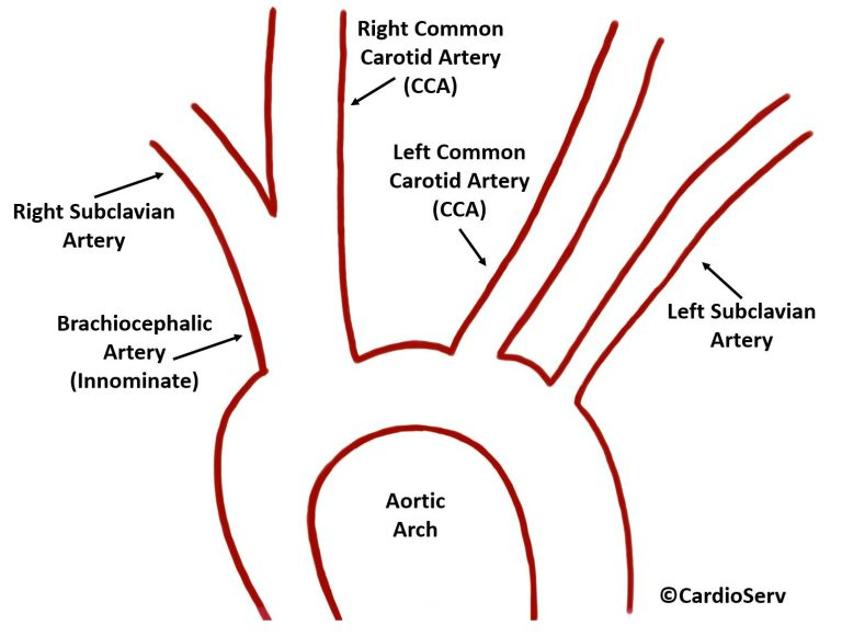
AORTIC ARCH
AORTIC ARCH BRANCHES:
- Brachiocephalic Artery (Innominate)
- Left Common Carotid Artery (CCA)
- Left Subclavian Artery
BRACHIOCEPHALIC ARTERY
- First branch off of aortic arch
- Supplies blood to right arm, neck, head
- Branches into:
- Right Common Carotid Artery (CCA)
- Right Subclavian Artery
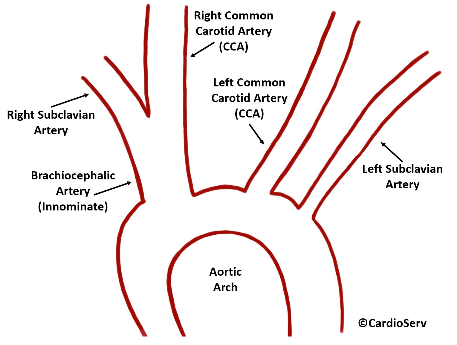
SUBCLAVIAN ARTERY
- Right subclavian artery branches off of brachiocephalic artery
- Left subclavian artery branches directly off aortic arch
- Major arteries of upper throax below clavicle
- Provides blood to arms
- Right subclavian artery: right arm
- Left subclavian artery: left arm
COMMON CAROTID ARTERY (CCA)
- Paired structure supplying neck & head with oxygenated blood
- Right CCA: branches off of brachiocelphalic artery
- Left CCA: branches directly off of aortic arch
- Move upwards on neck to level of thyroid cartilage
- Normal Diameter: 0.75 – 1.25 cm
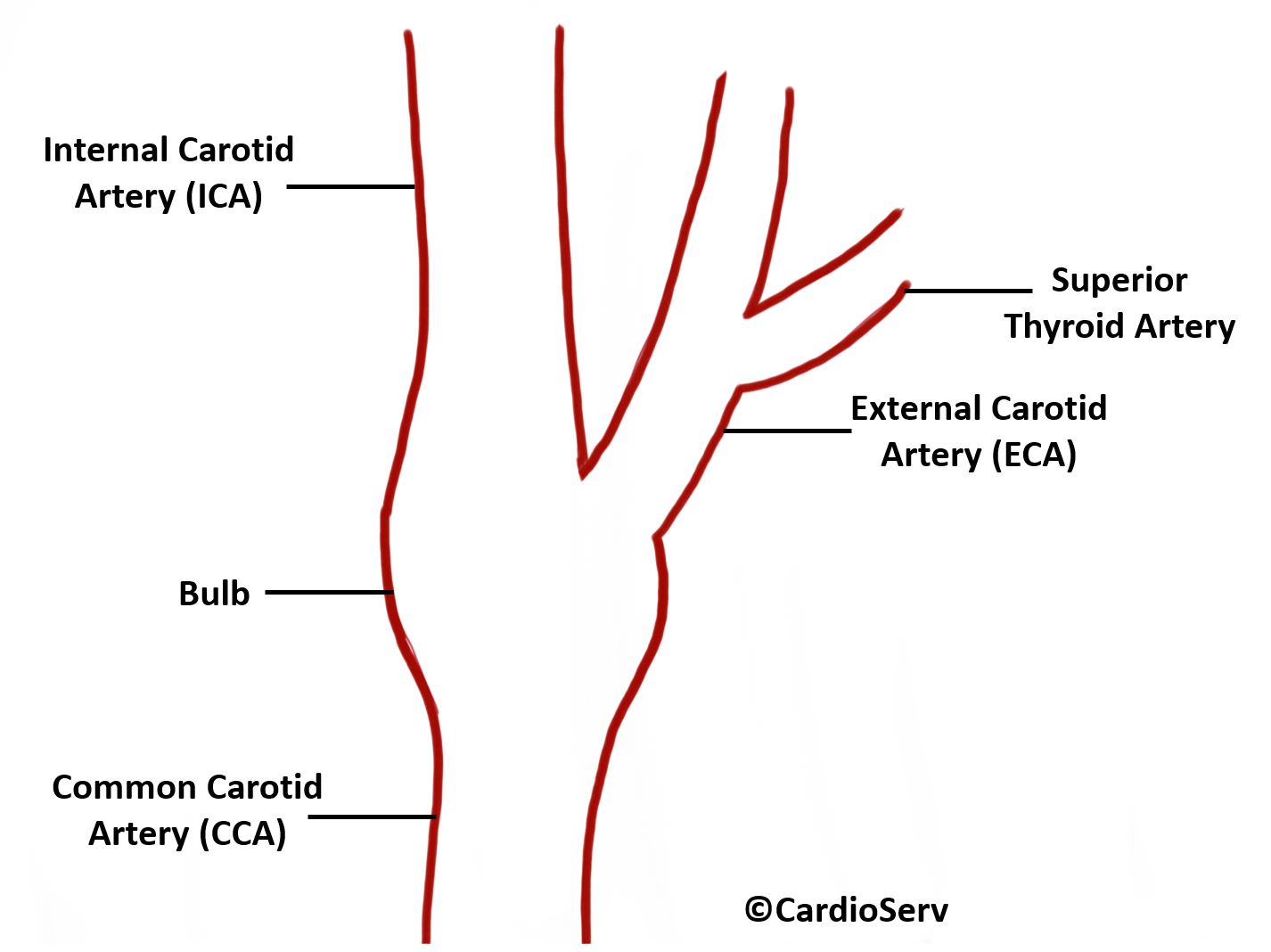
CAROTID ARTERY BIFURCATION
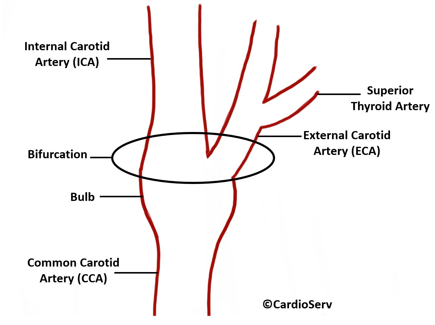
- Carotid Bulb: at level of bifurcation, the vessel will become enlarged
- Bifurcation: division of a vessel into multiple parts
- CCA Bifurcates into:
- Internal Carotid Artery (ICA)
- External Carotid Artery (ECA)
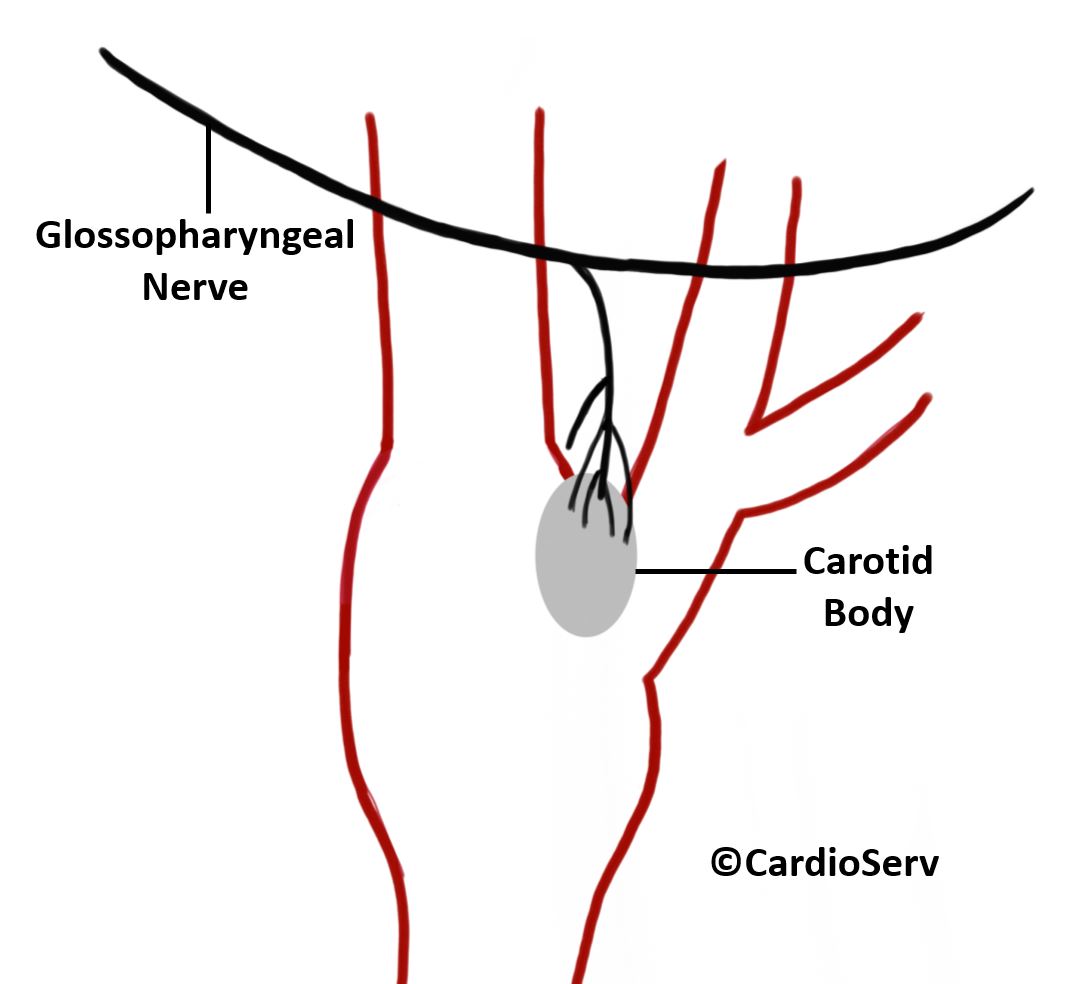
CAROTID BODY
- Small oval structure that sits behind bifurcation
- Function: small cluster of chemoreceptors- responds to oxygen (O2), carbon dioxide & pH levels in blood
- Glossopharyngeal Nerve: provides motor & sensory functions
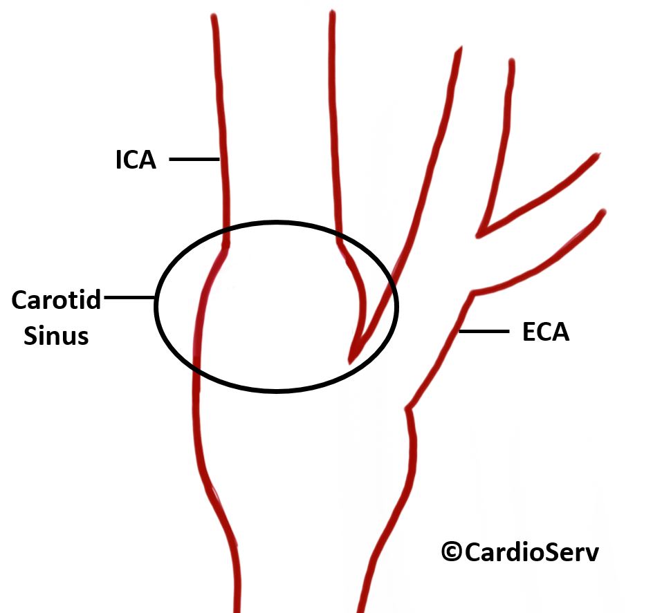
CAROTID SINUS
- Localized dilatation at origin of ICA & bifurcation of CCA
- Function: baroreceptor (pressure receptor)- regulates & maintains blood pressure
EXTERNAL CAROTID ARTERY (ECA)
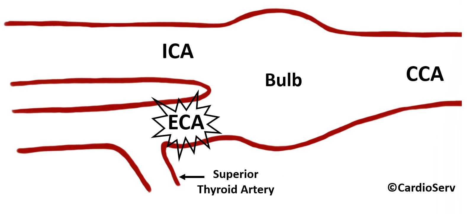
- Begins at bifurcation of CCA
- Travels upwards & terminates at superficial temporal artery (STA)
- Courses anterior & medial
- Smaller than ICA
- 1st major branch: superior thyroid artery
- Help distinguish between ECA & ICA
- Branches supply face, neck & head
- Develop collateral blood supply when carotid & vertebral disease present
- Normal Diameter: 0.25 – 0.70 cm
INTERNAL CAROTID ARTERY (ICA)
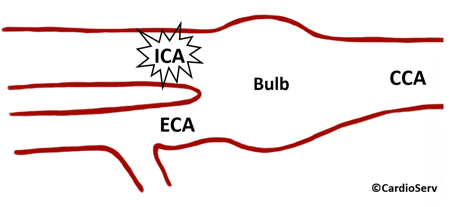
- Begins at bifurcation of CCA
- Travels upwards as single vessel until enters cranium (terminates)
- Courses posterior & lateral
- No branches within neck area
- Provides 75% blood supply to brain
- Normal Diameter: 0.5 – 1.0 cm
- Shape Distortions: due to embryologic, pathologic or aging
- Tortuous
- Kinked- associated with cerebral ischemia symptoms
- Coiled
SUMMARY
Review of basic anatomy is a helpful to in order to fully understand how to evaluate and assess for pathology!