Last Updated on November 18, 2025 by Don Gerig, RDCS
Now that diastology is a mandated part of the scanning protocol for all of our accreditation clients, we are starting to see more diastology measurement errors. We thought that sharing these errors may help others to avoid them. This week, we will review correct tissue Doppler measurement techniques while reviewing two common errors. These errors include:
- Cursor Placement
- Caliper Placement: Waveform Measurement
CORRECT TISSUE DOPPLER MEASUREMENTS (TDI)
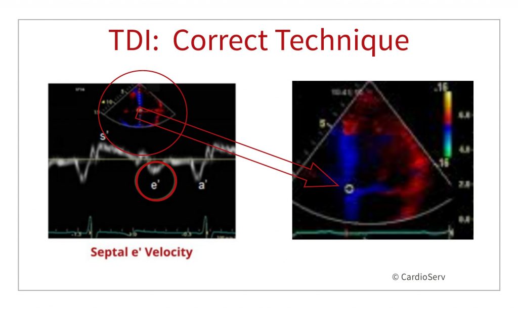
First, lets quickly review PW Tissue Doppler Imaging:
- TDI is one of the measurements needed to evaluate diastolic function and is used to estimate myocardial motion.
- We use TDI to measure the e’ velocity (cm/sec) in both the medial and lateral MV annulus locations.
- We use these measurements to calculate the average E/e’.
- Correct placement of the cursor should include the sample volume on the septal and/or lateral annulus of the mitral valve.
- Correct caliper placement includes measuring the e’ wave.
WHAT’S WRONG WITH THESE IMAGES?
Let’s review some sample images and see if you can identify the mistake.
Image 1
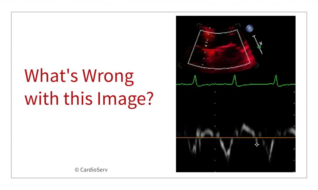
Did you notice the cursor placement on the septal annulus? You can see that the cursor over-shot the septal mitral valve annulus and is actually placed within the right heart. Incorrect cursor placement will greatly affect your Doppler readings. It’s important to take the time to correctly place your cursor.
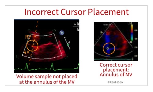
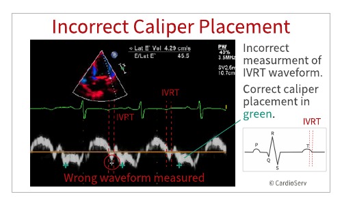
Image 2
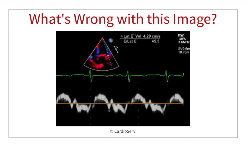
Did you notice the caliper placement on the tissue Doppler spectral waveform? The incorrect wave form has been measured. In this image there is a prominent IVRT waveform that was inadvertently measured as the e’ wave. An incorrect e’ will affect your E/e’ ratios.
SUMMARY
This week we reviewed two common mistakes seen with diastology measurements. Both cursor alignment and correct caliper placement will determine the value used for key measurements for assessing diastolic function. It is important to take the time to correctly measure these values.
Previous ‘What’s Wrong With This Image’ posts:
References
Seri, I., & Kleinman, C. S. (2019). Hemodynamics and cardiology(3rd ed.). Amsterdam: Elsevier. doi:https://www.sciencedirect.com/science/article/pii/B9780323533669000120
PAST ARTICLES ON DIASTOLOGY
- Understanding Diastology
- Correct Techniques to Acquire Diastology Measurements
- Specific Echo Parameters that Indicate Elevated LAP

Judith Buckland, MBA, RDCS, FASE
Stay connected: Facebook, Twitter, Instagram, LinkedIn


