Last Updated on November 18, 2025 by Don Gerig, RDCS
Better Education. Better Outcomes. Better Cardiology.
Welcome to the first post in our new Reader Q&A Series, where we take real questions from our audience and break them down into practical, easy-to-understand answers.
The Question
“Why does the flow convergence appear red on echocardiogram, even when the flow is away from the probe in the apical 4-chamber view?”
The Answer
At first glance, this seems counterintuitive. In the standard color Doppler map, red = toward the probe and blue = away from the probe. So if mitral regurgitation flow is away from the probe in A4C, shouldn’t the convergence zone be blue?
The key lies in color Doppler aliasing.
Understanding Color Doppler Aliasing
- Doppler ultrasound can only display flow velocities up to a certain threshold: the Nyquist limit (½ the pulse repetition frequency).
- When blood flow velocity exceeds this limit, the system “wraps around” and displays the flow in the opposite color.
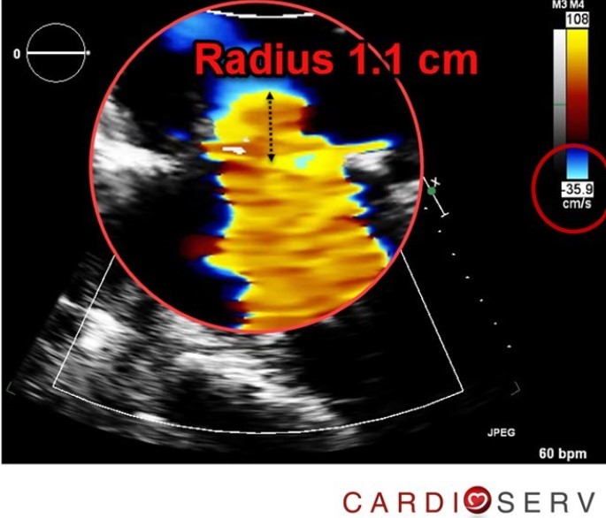
- In the flow convergence zone (PISA), blood accelerates rapidly toward the regurgitant orifice. The velocity near the valve is high enough to exceed the Nyquist limit → the color “aliases” from blue (true away flow) to red.
That’s why the classic flow convergence shell shows up as red and yellow, even though the flow is actually directed away from the probe.
Does Adjusting the Baseline Change This?
Yes!
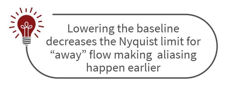
- Lowering the baseline decreases the Nyquist limit for “away” flow. This makes aliasing happen earlier, so the red and yellow hemisphere appears larger and easier to measure.
- Raising the baseline has the opposite effect: it reduces aliasing and keeps more of the convergence zone blue.
For PISA quantification, the baseline is typically shifted downward so the aliasing velocity is set around 30–40 cm/s. This produces a clear, measurable red hemisphere just proximal to the regurgitant orifice.
Quick Takeaway
Flow convergence looks red in A4C because of aliasing, not because the flow direction has changed. By adjusting the Doppler baseline, we can optimize the aliasing zone to make measurements more accurate.
✨ Have a question you’d like us to answer?
Drop it in the comments or send us a message. Your question might be featured in our next Reader Q&A Series blog!
DM us on social media! Facebook, Instagram or X (Twitter)
Related Articles:
Related online CME learning:
References
- Zoghbi WA, Adams D, Bonow RO, et al. Recommendations for Noninvasive Evaluation of Native Valvular Regurgitation. J Am Soc Echocardiogr. 2017 Apr;30(4):303–371.
- Otto CM, Pearlman AS. Textbook of Clinical Echocardiography. 6th Edition. Philadelphia, PA: Elsevier; 2018.
- Hoskins PR, Martin K, Thrush A. Diagnostic Ultrasound: Physics and Equipment. 3rd Edition. Cambridge University Press; 2019.