Last Updated on October 10, 2025 by Don Gerig, RDCS
One of the largest risk factors for cardiovascular disease today is hypertension. Not only that, it’s also one of the leading causes of death in the United States. The American Heart Association (AHA) emphasizes the importance of managing these risks early, especially by monitoring and controlling blood pressure.
When left untreated, hypertension can lead to hypertensive heart disease, including coronary artery disease (CAD), left ventricular hypertrophy (LVH), and hypertrophic cardiomyopathy (HCM). Echocardiography plays a pivotal role in detecting these changes, particularly through the evaluation of left ventricular mass and remodeling patterns.
In this article, we’ll explore the three main categories of LVH: concentric, eccentric, and concentric remodeling. Then we’ll translate what each those means for your daily echo practice.
What is Left Ventricular Hypertrophy (LVH)?
Left ventricular hypertrophy is a form of cardiac remodeling where the left ventricular wall thickens as a response to chronic increased pressure or volume overload. This thickening leads to an increase in LV mass (LVM) which can alter both systolic and diastolic function.
There are different sub-categories that fall into the diagnosis of LVH, which includes concentric, eccentric and concentric remodeling.
What Influences LVH Development?
There are several key factors that play a major role in determining the presence of LVH:
- Sex: Men typically have larger hearts than women.
- Age: Myocardial stiffness and wall thickness increase with age.
- Body Size: Athletes and obese patients often have larger hearts.
- Blood Pressure: Chronic hypertension drives concentric wall thickening.
- Heart Rate, Medications, and Diabetes: These can influence ventricular remodeling and hypertrophy patterns.
Key Point: Heart size is influenced by body size. always interpret LV dimensions and mass in context.
What Happens as the Heart Enlarges
As the ventricle adapts to chronic stress, wall stress increases, prompting the myocardium to thicken in order to normalize that stress.
Enlarged hearts: ↑ Wall stress → ↑ Wall thickness → ↑ LV cavity size (radius and volume)
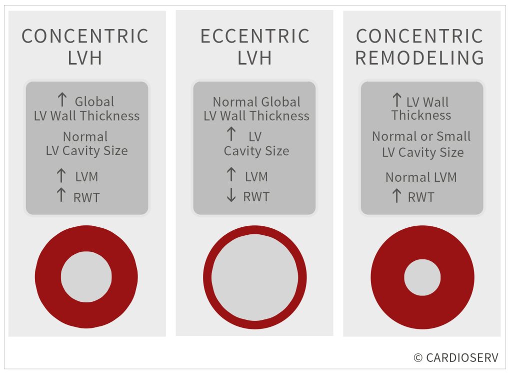
Concentric Left Ventricular Hypertrophy (LVH)
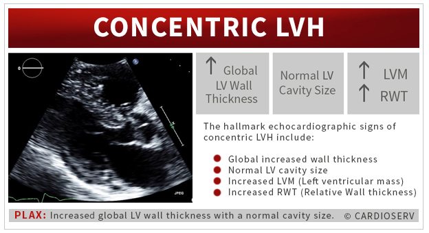
Concentric LVH is the result of the heart adapting to high systemic pressure overload caused by hypertension or other diseases such as aortic stenosis. Increased peripheral resistance forces the myocardium to thicken in order to normalize wall stress, a classic compensatory response to pressure.
This remodeling pattern affects both men and women across all ages and is associated with changes in LV geometry, diastolic filling, and myocardial strain.
Classic Signs of Concentric LVH on an Echocardiogram
- Increased global wall thickness
- Normal LV cavity size
- Increased left ventricular mass (LVM)
- Increased Relative Wall thickness (RWT)
Teaching Pearl: Concentric LVH = pressure overload pattern: thick walls, normal cavity, and increased wall-to-radius ratio.
Eccentric Left Ventricular Hypertrophy (LVH)
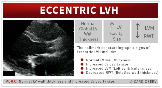
On the other hand, eccentric LVH develops in response to chronic volume overload, usually from valvular regurgitation or a high cardiac output state. In this setting, systemic pressures remain normal and peripheral resistance is not elevated, but the left ventricle dilates to accommodate the excess volume.
This category of patients will have changes in both diastolic function and the longitudinal and radial function. They will show to have either a low normal or a mildly impaired systolic function due to having chronic volume overload. The shape of the LV will change, becoming more spherical since the LV will be enlarged..
Classic Signs of Eccentric LVH on an Echocardiogram
- Normal LV wall thickness
- Increased LV cavity size
- Increased LVM (Left ventricular mass)
- Decreased RWT (Relative Wall thickness)
Teaching Pearl: Eccentric LVH = volume overload pattern: enlarged chamber, normal wall thickness, and low RWT.
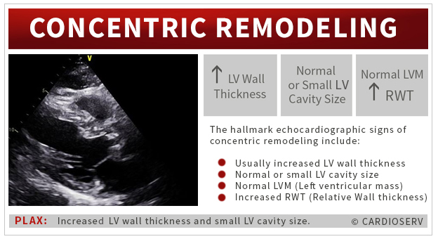
Concentric Remodeling
Concentric remodeling represents a late-stage adaptive response to chronic stress on the left ventricle, oftentimes a result of longstanding hypertension, chronic pressure or volume overload, or prior myocardial infarction (MI).
In this stage, the ventricle begins to lose its normal elliptical (bullet-shaped) geometry and becomes more rounded. This structural change is accompanied by progressive diastolic dysfunction and gradual decline in radial and longitudinal function, signaling early systolic impairment.
Concentric remodeling is the late stage response to LV hypertrophy; caused by either chronic pressure, volume overload or a MI (which is commonly associated with CAD, but can be due to longstanding hypertension, especially untreated). Concentric remodeling will demonstrate systolic dysfunction along with changes in the LV geometry. The shape of the LV changes and becomes more rounded, rather than bullet shaped. You will notice the rapid degradation of diastolic dysfunction and loss of radial and longitudinal function.
Classic Signs of Concentric Remodeling on an Echocardiogram
- Usually increased LV wall thickness
- Normal or small LV cavity size
- Normal LVM (Left ventricular mass)
- Increased RWT (Relative Wall thickness)
Final Thoughts and Next Steps
Our goal this week was to provide you with a better understanding of the pathophysiology behind the various categories of LVH. This table will provide you with a better understanding of the echocardiographic findings found in the subcategories of left ventricular hypertrophy (LVH). We discussed how LV mass (LVM) and relative wall thickness (RWT) plays a role in identifying the categories of LVH. But what is LVM and RWT?? How do we obtain them? In the second part of our LVH blog series, we will provide step-by step instructions on how to obtain these values.
REFERENCES
- Alam, S. (n.d.). The Edinburgh Cardiology Imaging Website. Retrieved March 01, 2017, from http://webservice1.mvm.ed.ac.uk/imaging/demo/cases/cardiac-amyloid-echo.html
- Marwick, T. H., PhD, Gillebert, T. C., MD, & G. A., MD. (2015). Recommendations on the Use of Echocardiography in Adult Hypertension. JASE,28(7), 1-12. Retrieved March 1, 2017, from http://asecho.org/wordpress/wp-content/uploads/2015/07/2015_Hypertension.pdf
- Lang RM, Badano LP, Mor-Avi V, et al. Recommendations for Cardiac Chamber Quantification by Echocardiography in Adults: An Update from the American Society of Echocardiography and the European Association of Cardiovascular Imaging. J Am Soc Echocardiogr. 2015;28(1):1–39.e14. DOI:10.1016/j.echo.2014.10.003


