Last Updated on November 19, 2025 by Judith Buckland, MBA, RDCS, FASE
In earlier posts we have reviewed RV remodeling in pulmonary hypertension. If adaptive and maladaptive remodeling describe how the RV looks and behaves, RV–PA coupling describes how well it works.
The concept of RV–PA coupling and uncoupling can sound intimidating, but it’s really about how well the right ventricle (RV) and the pulmonary arteries “work together”.
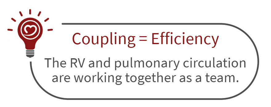
Coupling is the measure of teamwork between the right ventricle and the pulmonary circulation—how efficiently the RV converts its contraction into blood flow through the lungs.
- When coupling is intact, the RV and pulmonary arteries are perfectly matched, maintaining cardiac output with minimal wasted energy.
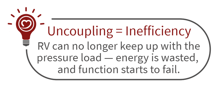
- When that balance breaks down, uncoupling begins—the hallmark of declining efficiency and the first signal that the RV is losing ground against rising pulmonary pressures.
🟢 What Is RV–PA Coupling? When the RV and Lungs are in Sync
When the RV and lungs are in sync, the RV pumps just the right amount of force to push blood into the pulmonary arteries efficiently — not too hard, not too weak.
That perfect balance is called coupling.
- The RV muscle is strong enough to handle the pressure in the lungs.
- Blood flow is smooth and efficient.
- The heart doesn’t waste energy — it’s working smart, not hard.
- On echo, the RV still looks strong and coordinated (good TAPSE, FAC, strain, and normal shape).
RV–PA coupling describes the mechanical efficiency between the RV’s contractile performance and the pulmonary circulation’s afterload. This relationship is quantified as the ratio of end-systolic elastance (Ees) to arterial elastance (Ea)
🔴 What Causes RV–PA Uncoupling? When the Teamwork Breaks Down
Over time, as pulmonary pressures rise, the RV has to push harder and harder.
At first, the RV keeps up by thickening its walls (adaptive hypertrophy). But eventually, it can not generate enough strength to match the rising pressure — it tires out.
That’s when uncoupling happens.
- The RV and pulmonary arteries are out of sync.
- The RV can’t push blood effectively against the high resistance.
- Blood backs up into the right atrium and body (legs, liver, abdomen).
- The RV becomes enlarged, weaker, and less efficient — like an athlete who’s overtrained and exhausted.
- On echo, you’ll see lower TAPSE, FAC, and strain, plus septal flattening (the “D-shaped LV”).
RV–PA Coupling: The Efficiency Connection
RV–PA coupling describes the mechanical efficiency between the RV’s contractile performance and the pulmonary circulation’s afterload.
This relationship is quantified as the ratio of end-systolic elastance (Ees) to arterial elastance (Ea):
Ees/Ea ≈ 1.5–2.0 = optimal coupling and efficient energy transfer.
What Ees/Ea Represents
- Ees (end-systolic elastance) = the contractility of the right ventricle — how strongly it squeezes, independent of load.
- Ea (arterial elastance) = the afterload faced by the RV — how “stiff” or “resistive” the pulmonary arteries are.
- Their ratio (Ees/Ea) reflects how efficiently the RV converts contractile energy into forward flow through the pulmonary circulation.
- Normal / Coupled: Ees/Ea ≈ 1.5–2.0
- Uncoupled: Ees/Ea < 1.0
The true way to quantify Ees and Ea is via a right heart catheterization, but it’s mostly done in research, not routine practice.
Echo Surrogates (Clinical Practice)
Since calculating Ees/Ea via cath is not feasible day to day, echo provides validated surrogate ratios that parallel Ees/Ea efficiency.
1. TAPSE/sPAP ratio
- TAPSE (mm) = longitudinal shortening of the RV free wall.
- sPAP (mmHg) = systolic pulmonary artery pressure (from TR jet).
- Interpretation:
- ≥0.31 mm/mmHg → Coupled
- <0.31 mm/mmHg → Uncoupled / early RV failure
- Simple, reproducible, and widely used clinically.
2. RV Free Wall Strain / sPAP
- Strain from speckle tracking (longitudinal % shortening).
- Cutoff: ratio < -0.44 %/mmHg suggests uncoupling.
3. FAC/sPAP or S′/sPAP
- Variants for labs that measure different RV function indices.
- All express RV contractile performance relative to pressure load.
Conclusion
RV–PA coupling turns the abstract idea of “RV function” into something measurable and actionable.
- A coupled system reflects efficiency and reserve—the RV and pulmonary circulation moving in harmony.
- Uncoupling marks the breaking point, when the RV’s efforts no longer meet the demands of the pulmonary circuit.
By understanding and tracking this relationship, clinicians can detect failure earlier, intervene sooner, and preserve right-heart performance before structural damage becomes irreversible.
Related Articles and Resources
- Pulmonary Hypertension Case Study Presentation
- Understanding Pulmonary Hypertension in Echo
- Mastering TAPSE in 2025: Graded Severity, Pitfalls, and Best Practices
- 2025 ASE Right Heart Guidelines

Judith Buckland, MBA, RDCS, FASE
Connect on LinkedIn