Last Updated on November 18, 2025 by Don Gerig, RDCS
New Aortic Valve Blog Series
This next blog series will cover the different aortic cusp variants and how they present in echocardiography. This week we will begin our review of the unicuspid aortic valve and we will cover the bicuspid and quadricuspid AV in future blogs.
If you are in need of CMEs we offer a 5.0 Cat 1, AMA CME course that provides a detailed look at Aortic Valve Anatomy and Cusp Variants in Echocardiography including the unicuspid, bicuspid, tricuspid and quadricuspid aortic valve.
Unicuspid Aortic Valve (UAV)
- UAV has only one functional leaflet
- Rare congenital disorder that affects an estimated 0.02% of the adult population.
- Prevalence of unicuspid aortic valve is higher in patients who undergo valve replacement for aortic stenosis, ranging from 4–6%
- Difficult to distinguish from bicuspid aortic valve (BAV)
- UAV share valve pathology similar to BAV, such as:
- raphe
- leaflet fusion
- aortic stenosis and aortic regurgitation
- ascending aortic dilatation
- UAV consists of a single commissural zone of attachment or absence of a commissural attachment
- In adults the unicuspid aortic valve is mainly unicommissural and the free edge of the aortic valve leaflet arises from a single aortic attachment that encircles the entire orifice
- This gives the unicuspid aortic valve the “circular” appearance on echo
Cause of Unicuspid Aortic Valve (UAV):
- A rare congenital disorder that affects an estimated 0.02% of the adult population.
- It may be associated with genetic syndromes or isolated as a congenital anomaly.
Mechanism of Unicuspid Aortic Valve (UAV):
- The valve’s structure leads to severe hemodynamic stress, causing rapid degeneration and severe aortic stenosis early in life.
- The lack of normal valve opening causes turbulent flow across the valve, leading to calcification and fibrosis of the solitary cusp.
Anatomical Features of the Unicuspid Aortic Valve:
- UAV consists of a single commissural zone of attachment or absence of a commissural attachment
- The valve has a single cusp that opens in a slit-like orifice during systole.
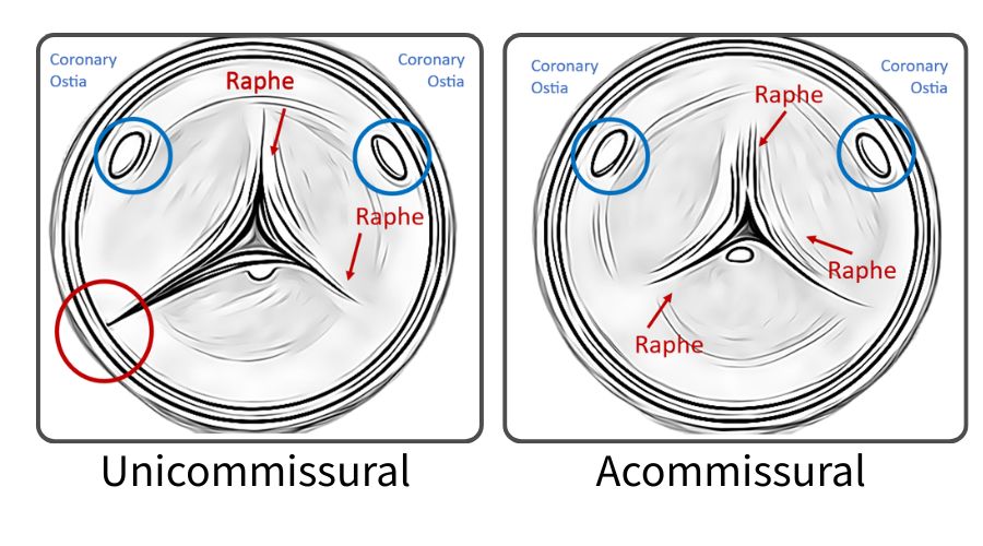
Unicuspid AVs can be further categorized into two types: Unicommissural and Anicommissural.
Unicommissural Valve:
- There is one lateral commissural attachment to the aorta at the level of the orifice
- Appears as a slit shaped structure
- Patients typically present in the 4th to 6th decade of life
- Rarely can present at infancy.

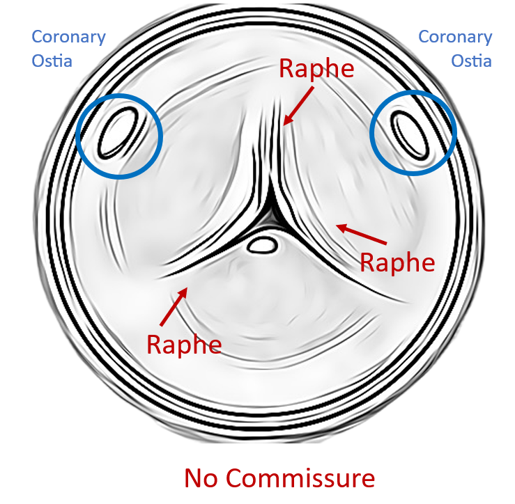
Acommissural Valves:
- There are no commissures or lateral attachments to the aorta
- Orifice is very small and appears as a pinhole on imaging
- Patient have symptomatic severe aortic stenosis at birth or infancy
Progression:
- Rapid progression to severe stenosis occurs in early adulthood or adolescence.
- Often leads to early valve replacement, typically in the teenage years or early adulthood due to severe obstruction of blood flow.
Other Features:
- The unicuspid valve is prone to aortic aneurysm formation due to the abnormal stress placed on the aortic root and ascending aorta.
Age Group:
Infants, children, or adolescents often present with severe stenosis or regurgitation, much earlier than bicuspid or trileaflet aortic stenosis.
Next Month
Next week we will review how the unicuspid AV presents in echocardiography. Sign up to recieve our blogs via email.
Related Blogs:
- Aortic Valve Anatomy
- Aortic Stenosis and Mismatch Values
- Top 5 Factors to Consider when Evaluating AS
Check out our full online learning platform at cme.cardioserv.net!