Written by Yvonne Prince ACS, RDCS, RVT, RDMS, FASE
In an early blog, Abdominal Ultrasound for Echocardiographers: Part 1, we reviewed some basic tips for echocardiographers scanning the abdomen. We reviewed artifacts, image orientation and patient positioning. This week we will provide you with 6 steps to successfully identify the aorta and...
Written by Yvonne Prince ACS, RDCS, RVT, RDMS, FASE
Branches of the Aorta
Earlier, I mentioned the superior mesenteric artery (SMA). From a longitudinal approach, it is seen rising anteriorly and then turning and coursing distally, maintaining a parallel course to the aorta. The SMA is the second branch of the abdominal aorta, and the celiac axis...
Written by Andrea Fields MHA, RDCS
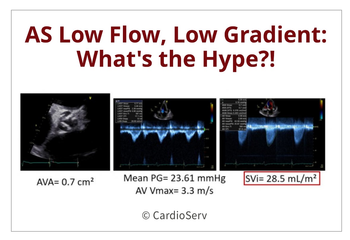
We've all been in this situation.... our patient appears to have a very tight, calcified aortic valve which visually appears to be moderate to severe aortic stenosis. As we scan through our protocol, we obtain a peak aortic valve velocity of 3.3 m/s and mean pressure gradient (PG) of 23.6 mmHg. The...
Written by Yvonne Prince ACS, RDCS, RVT, RDMS, FASE
How often have you found yourself “in over your head” in the abdomen when trying to image the IVC and abdominal aorta? Is imaging the abdominal aorta part of your echo protocol? It is not uncommon for a patient to receive an abdominal ultrasound because the echo findings mentioned the...
Written by Andrea Fields MHA, RDCS
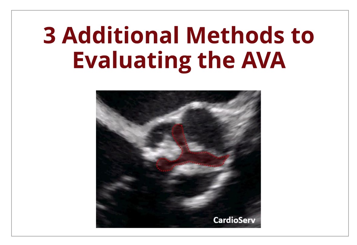
Last week we broke down the continuity equation to evaluate the severity of aortic stenosis. This week, we are going to discuss other methods that can be used to evaluate the severity of aortic stenosis. This blog will cover the following methods:
AVA Planimetry
Indexed Continuity...