Written by Yvonne Prince ACS, RDCS, RVT, RDMS, FASE
Branches of the Aorta
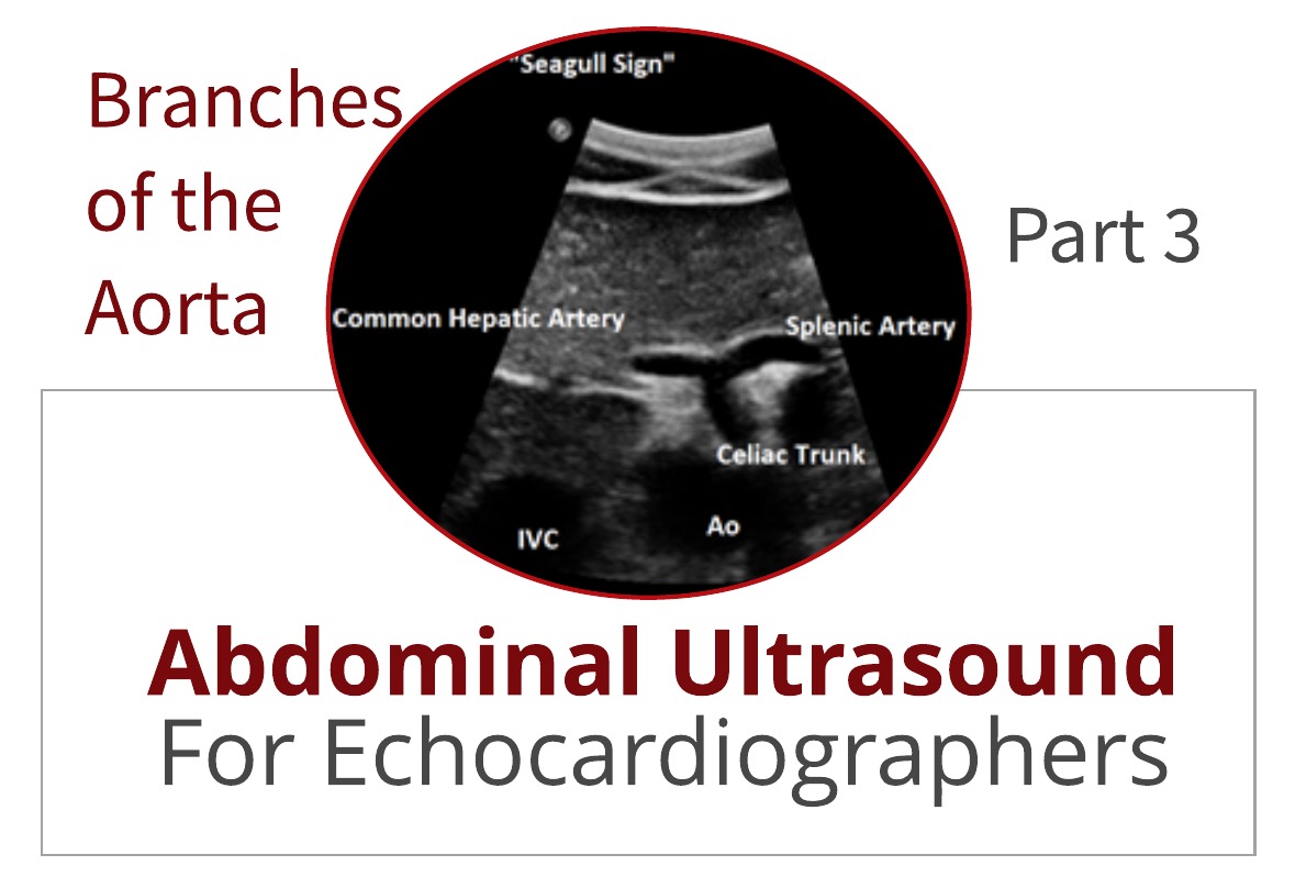
Earlier, I mentioned the superior mesenteric artery (SMA). From a longitudinal approach, it is seen rising anteriorly and then turning and coursing distally, maintaining a parallel course to the aorta. The SMA is the second branch of the abdominal aorta, and the celiac axis...
Written by Yvonne Prince ACS, RDCS, RVT, RDMS, FASE
If you have been following along with Parts 1 and 2 of this blog series, you have already learned how important landmarks are in scanning the abdominal vasculature. We provided a step-by-step method for identifying and imaging the abdominal aorta. This week in Part 3, we will discuss tips and...
Written by Yvonne Prince ACS, RDCS, RVT, RDMS, FASE
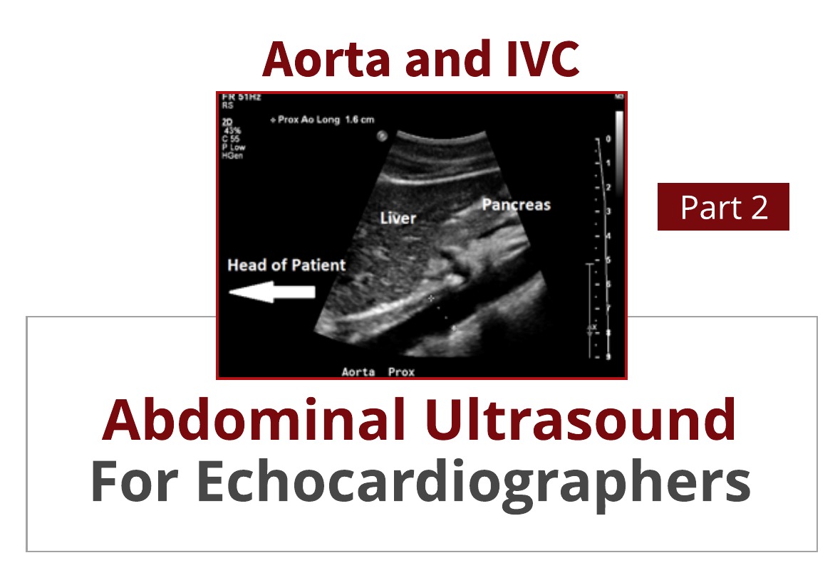
In an early blog, Abdominal Ultrasound for Echocardiographers: Part 1, we reviewed some basic tips for echocardiographers scanning the abdomen. We reviewed artifacts, image orientation and patient positioning. This week we will provide you with 6 steps to successfully identify the aorta and...
Written by Yvonne Prince ACS, RDCS, RVT, RDMS, FASE
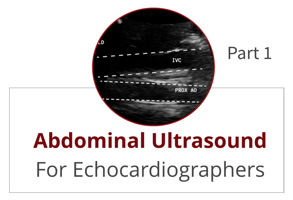
How often have you found yourself “in over your head” in the abdomen when trying to image the IVC and abdominal aorta? Is imaging the abdominal aorta part of your echo protocol? It is not uncommon for a patient to receive an abdominal ultrasound because the echo findings mentioned the presence...
 Cardioserv Blog