Written by Andrea Fields MHA, RDCS
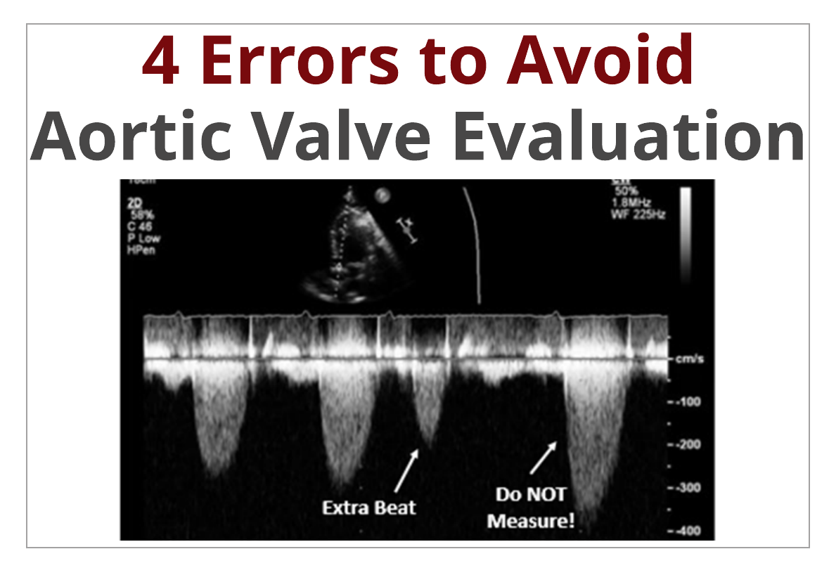
Evaluating the aortic valve is a routine part of an echocardiogram. There are many methods we use to determine the structure and function of the valve, including 2D, color Doppler and a combination of pulsed & continuous-wave Doppler. With short examination times being a common challenge, it...
Written by Judith Buckland, MBA, RDCS, FASE
As CardioServ continues our Educational World Tour, our next destination took us to Mumbai, India! Here, we volunteered to speak at the Essentials of Echocardiography conference, which was held at Sir H.N. Reliance Foundation Hospital on January 7, 2018. This blog will describe our experience...
Written by Andrea Fields MHA, RDCS
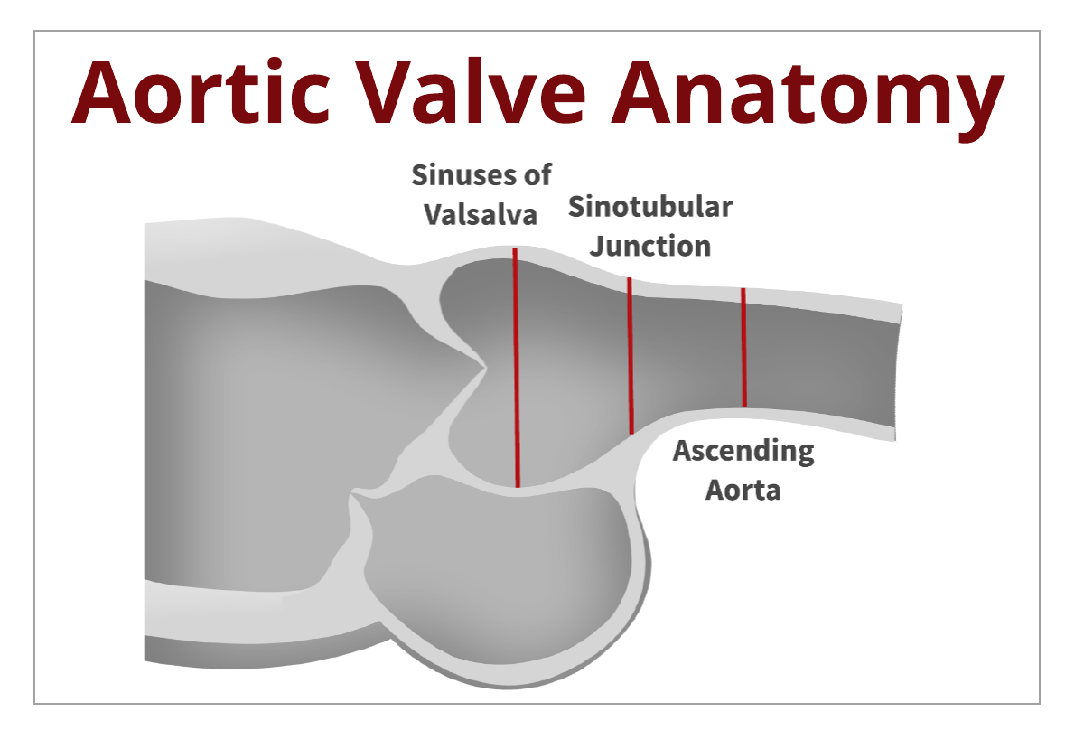
We evaluate the aortic valve for pathology on every patient we scan. It's easy to forget the basic concept of anatomy and the functions each part of the valve plays in opening & closing of the leaflets. This blog is going to be a refresher and cover the basic anatomy of the aortic...
Written by Andrea Fields MHA, RDCS
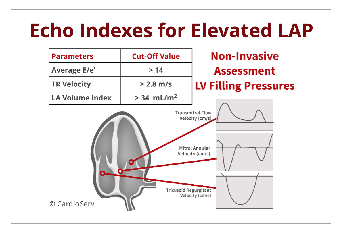
Last week, we discussed change in transmitral filling patterns as the severity of diastolic dysfunction progresses. When we evaluate for diastolic dysfunction, our goal is to determine:
Grade of diastolic dysfunction
Presence or absence of elevated filling pressures
Determining LV...
Written by Andrea Fields MHA, RDCS
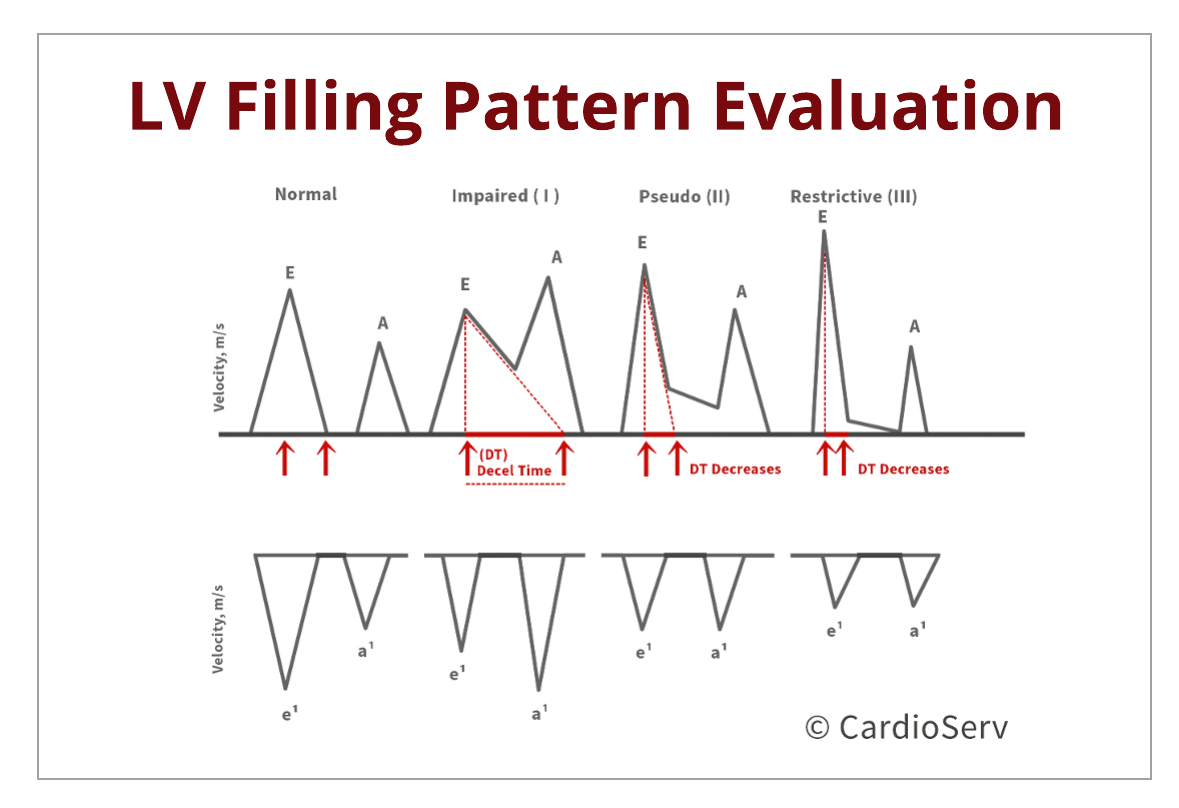
Let's discuss the transmitral filling patterns as the severity of diastolic dysfunction progresses. The transmitral pressure gradients are reflective upon the E & A-wave velocities. The waveforms are influenced by the ability of the ventricle to relax and the compliance of the LA to generate a...
 Cardioserv Blog