Written by Judith Buckland, MBA, RDCS, FASE and Andrea Fields, MHA, RDCS
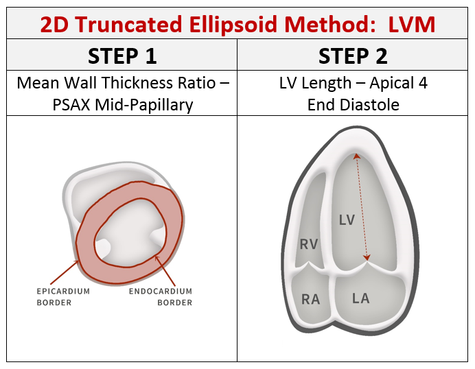
In earlier blogs we discussed calculating the left ventricular mass (LVM) via the 2D Linear Cubed Method with assumes an ellipse shape with a major/minor axis ratio of 2:1. We received a follow up question from a reader: “Can you please provide the reference for NOT using cube method...
Written by Judith Buckland, RDCS, FASE, MBA and Andrea Fields, MHA, RDCS
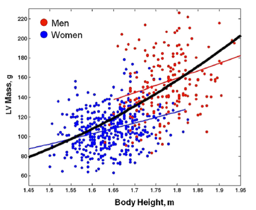
We recently posted a 2-part blog series on how to calculate LV Mass (LVM) and one of our readers provided some great feedback regarding the impact of height on LVM. We thought this reader’s comment was worth sharing and answering.
Reader: Using body surface area to normalize LVM will...
Written by Andrea Fields, MHA, RDCS
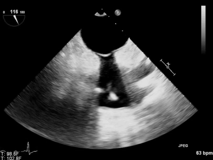
Name that Pathology!
Modality: Adult Echo TEE
Review the following images and see if you know what pathology is present!
TEE images of LA and RA
Color Doppler over the atrial septum demonstrating PFO.
TEE image of IAS
Answer: Lipomatous Atrial Septal Hypertrophy (LASH)
What is...
Written by Judith Buckland, RDCS, FASE, MBA
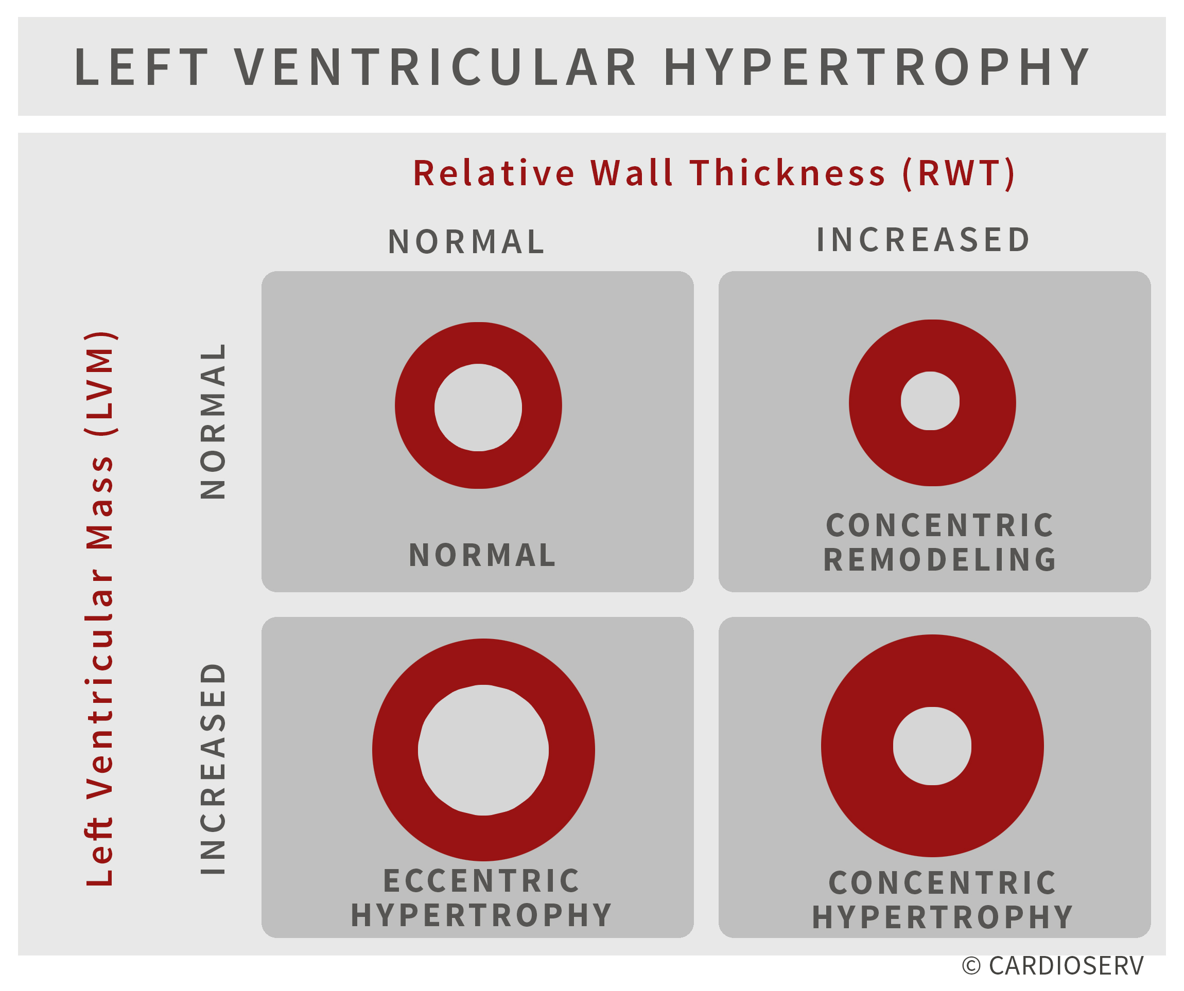
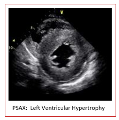
As diagnostic imaging professionals, we often perform echocardiograms on patients with hypertension to monitor the thickness, strength and wall motion of the heart. Last week we launched our two-part blog on left ventricular hypertrophy (LVH). In part one we explained the pathophysiology behind the...
Written by Judith Buckland, RDCS, FASE, MBA and Andrea Fields, MHA, RDCS
Last month was designated American Heart Month to raise awareness of cardiovascular disease, the leading fatality of Americans. AHA encourages our patients to take control over understanding their risk factors of heart disease including knowing their numbers related to blood pressure. Untreated...
 Cardioserv Blog