fbpx
Written by Andrea Fields MHA, RDCS

8 Tips to Correct RV Function Assessment with TAPSE & S’ Wave

We dedicated the month of April to learning about ways to implement right heart quantification into our routine exams.  This month, we are going to continue that journey and discuss ways to evaluate the function of the right heart!

RV systolic function should be assessed by at least one or combination of the following: FAC, S’ Wave, TAPSE or RIMP.” –ASE JASE 2015

With that in mind, let’s jump into how to perform these!

RV Systolic Function

The ASE explained 4 methods to quantify RV systolic function:

  1. TAPSE
  2. S’ Wave
  3. FAC
  4. RIMP

This week we are going to discuss how to perform TAPSE & S’ Wave measurements.

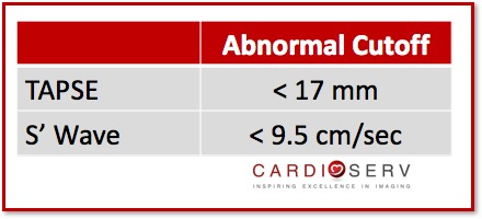
The reference values for these parameters do NOT provide a mild, moderate or severe value range. They do not take BSA, height or gender into consideration either.  ASE provides us with normal and abnormal values.

Imaging Window

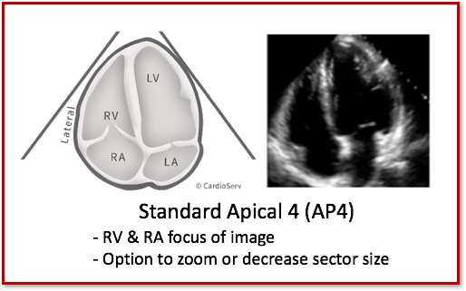
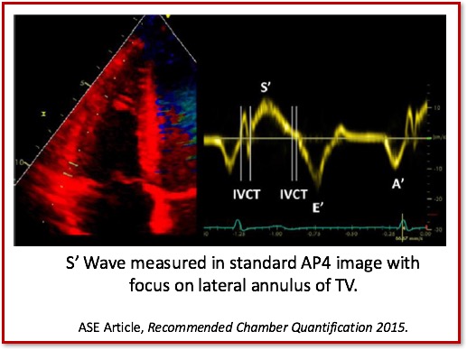
The ASE suggest using the standard AP4 window when performing RV function assessment.

Standard Apical 4 Chamber (AP4)

Standard AP4

Right Ventricle Function Assessment

TAPSE: Tricuspid Annular Plane Systolic Excursion

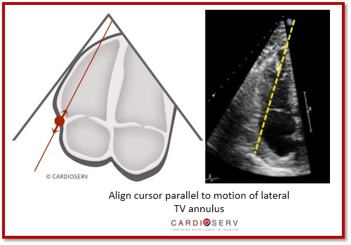
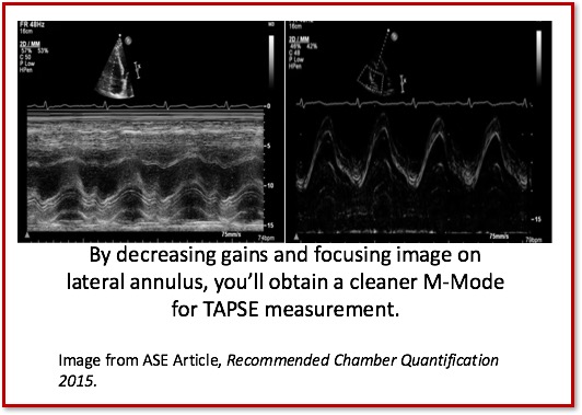
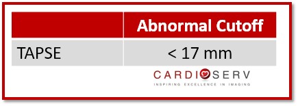
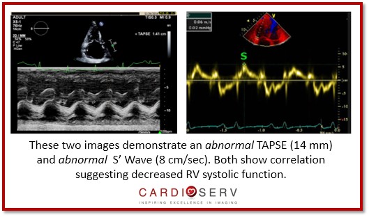
TAPSE is a linear M-Mode measurement of the RV longitudinal function.

  • Wide Spread
  • Established Prognostic Value
  • Validated with Global RV Function

How to Perform TAPSE Measurement

  • Align M-Mode cursor parallel to motion of lateral TV annulus**
    • Annulus should be moving upwards towards the apex during systole

Cursor Alignment

  • Decrease gains to avoid measuring ‘noise artifact’
  • M-Mode Speed: Medium-Fast
  • Look for a consistent signal throughout systole and diastole
  • Identify maximum systolic and diastolic excursion of annulus motion
  • Measure VERTICALLY via leading-edge to leading-edge method

TAPSE Gain Control

TAPSE Distance

Challenges/Limitations to linear measurements:

  1. Angle Dependency
  2. Easy to over and under-estimate RV function

Reference Values

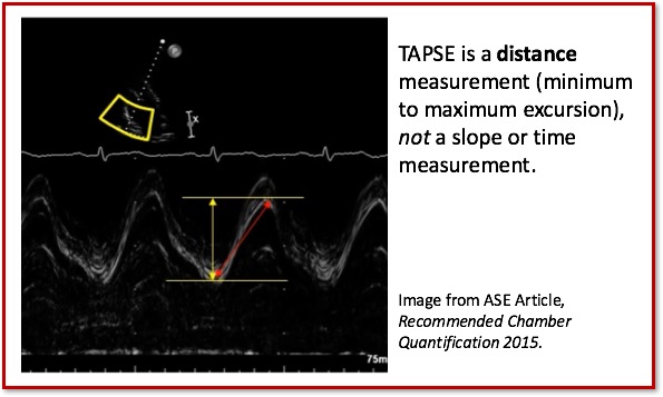
There were changes to the cutoff value for TAPSE in the latest ASE guidelines and ASE acknowledged that although there may be minor variations in TAPSE values according to gender and BSA that generally a TAPSE value less than 17mm is highly suggestive of RV dysfunction.

TAPSE Value

 S’ Wave: TDI-Derived Tricuspid Lateral Annular Systolic Velocity

Peak systolic velocity of the tricuspid annulus by Pulsed Wave TDI (cm/sec).

  • Good indicator of basal (lateral) free wall function
  • Easy to perform & reproduce
  • Established prognostic value
  • Similar concept to TAPSE

How to perform S’ Wave Measurement

  • Place cursor over lateral annulus of tricuspid valve
  • Pulse Wave Doppler and TDI
  • Decrease gains to avoid measuring ‘noise artifact’
  • Identify maximum systolic velocity ABOVE the baseline—‘S’ Wave’

S Wave

Challenges/Limitations to measurement:

  1. Angle Dependent
  2. Not fully representative of global RV function
  3. Easy to underestimate if Doppler signal is not parallel to annulus

Reference Values

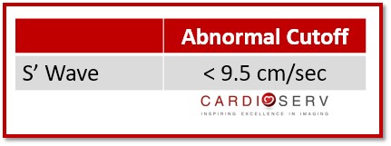
Again, there were minor changes to the S’ reference range with the latest ASE guideline update.  Just like TAPSE there are no values based upon mild, moderate or severe.  The reference range provided is a cutoff range with a S’ value less than 9.5 cm/sec indicating RV dysfunction.

S' Wave Value

**HELPFUL TIP:

It is VITAL that we make sure we obtain both TAPSE and S’ Wave values to correlate together. If you are getting an abnormal TAPSE, then you need to make sure your S’ Wave is demonstrating an abnormal value as well.

Here is an easy reference chart to refer to in your scanning laboratory:

Abnormal Values


TAPSE, S' Wave Abnormal

8 Tips to Measure TAPSE & S’ Wave

  1. Use the standard AP4 view
  2. Utilize chamber focused imaging: we are only interested in the RV & RA!
  3. Make cursor parallel to motion of annulus!
  4. Use gain controls to optimize image
  5. Look for consistent sign for accurate measurement
  6. Adjust sweep speed to elongate waveforms
  7. Make sure TAPSE & S’ Wave correspond correctly to each other! Either both abnormal or both normal!
  8. Implement both measurements into your normal scanning routine!

Conclusion

The ASE provides us with four great methods to use for evaluating the RV function. If you’re not using any method to quantify right heart function, today is the day to start! We at CardioServ, suggest using both TAPSE and S’ Wave. If you are only limited to one, S’ Wave is easily obtained and more reproducible. If you do get an abnormal S’ Wave, it would be idea to also perform a TAPSE as an additional method to proving your abnormal value. Next week, we will discuss two other methods [RIMP & FAC] we can use for evaluating right sided function!

Want a CME for reading this blog? Click below to access CardioServ’s online CME platform offering Cat. 1 AMA CMEs!

Access CMEs


Andrea Fields MHA, RDCS, Cardiac Clinical Director

Connect with me on LinkedIn


References

Lang, R. M., MD, Badano, L. P., MD, & Mor-Avi, V., PhD. (2015). Recommendations for Cardiac Chamber Quantification by Echocardiography in Adults: An Update from the American Society of Echocardiography and the European Association of Cardiovascular Imaging. JASE, 28(1), 1-53. Retrieved March 1, 2017, from http://asecho.org/wordpress/wp-content/uploads/2015/01/ChamberQuantification2015.pdf


adult echoASE guidelinescardiac chamber quantificationcmeechoechocardiographyright ventricleRV FunctionRV QuantificationS' WaveTAPSEtechnical measure

Our Services

LET US KNOW WHAT YOU THINK...

Cuong
Thank you! It is useful for me!
Eric
Excellent and very succinct.
Meena Sumathy
Excellent article on how to measure RV function. But often TAPSE is unreliable in significant tricuspid regurgitation ( as its load dependent). TDI value at lateral annulus is less load dependent ( but more angle dependent) and may have limitations with tricuspid valve pathologies. That makes both unreliable for assessing RV function in the above setting. Is RV strain the best method in these situation.
Steve
Meena, Although true, we should perform all measurements available to us so the reader can better interpret/diagnose. In such an instance, I would also perform FAC, Strain, and a 3-D assessment, if available.
Phillip D'Amato
Excellent article on the importance of proper right ventricle quantification.TAPSE is vital to proper analysis of right ventricle function.
Sarat
Good description
Krishnan
Great
Tayler
Nice
H. M. Ismail
Good work to keep up-to-date information.
Angela Bays
Very helpful. It's great to see things like this broken down.
Dawid de Wet
Thank you,very helpful.
drsalmankhan
informative
Abbas
Thank you for this nice data
Abbas
Thank you
Mohamed
Good
satish tiwari
very helpful
dr sirajur rahman
excellent for learning echo
Bao Tran
Good, easy to practice.
Javier Osorio
excelente
Cheryl Semrinec
Love what I seen!! Keep up the good work!
Khan
Thank you.
JAVIER OSORIO
Muy didactico, gracias.
Nilofer Ali
Thank you. It was helpful.
dr ajay walimbe
CAn TAPSE be used as parameter for fluid responsiveness or diagnosisng hypovolemia early.thanks waiting for reply
Azair
Thank you very much
DR KESTER C EFUGH
Thanks for the article.
Kimari Joseph
simple and clear,very helpful,i was not using these two methods, i will start now,thanks
Talal Da
Awesome video thank you so much !!

Your email will not be displayed.

Required *

Blog
Sign Up

Accreditation
services

Accreditation Help
Online Echo CMEs
Echo Accreditation Software
Free
Consultation

Search
for

Blog
Categories

Blog
Archive

Tags
Used

Show tags
Need Help? Share