Written by Yvonne Prince ACS, RDCS, RVT, RDMS, FASE
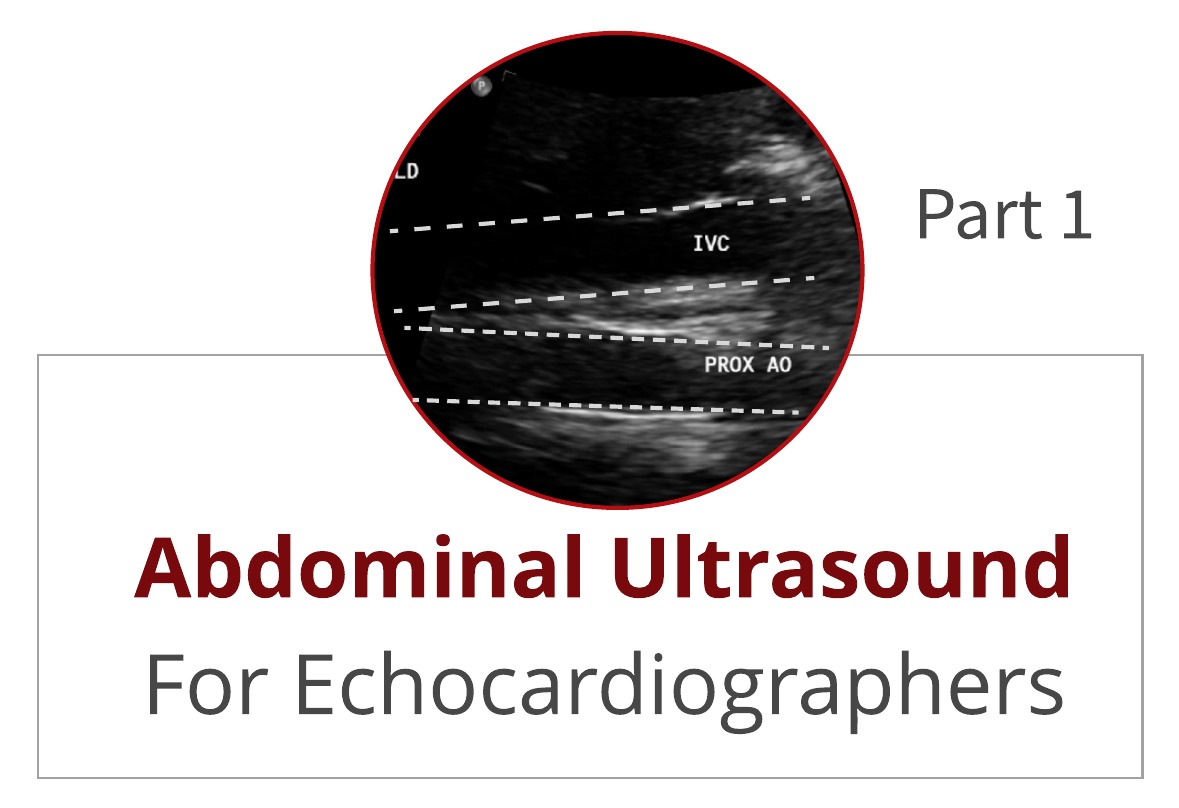
In an early blog, Abdominal Ultrasound for Echocardiographers: Part 1, we reviewed some basic tips for echocardiographers scanning the abdomen. We reviewed artifacts, image orientation and patient positioning. This week we will provide you with 6 steps to successfully identify the aorta and...
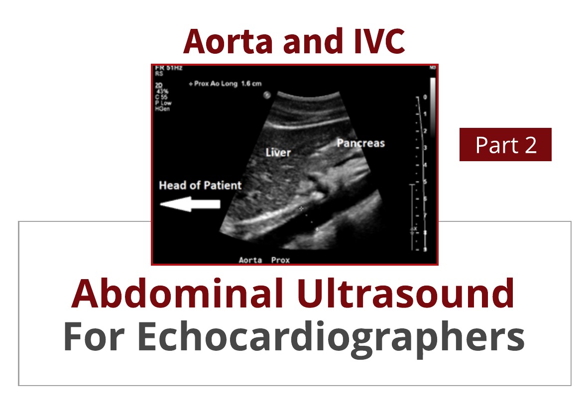
Written by Yvonne Prince ACS, RDCS, RVT, RDMS, FASE
How often have you found yourself “in over your head” in the abdomen when trying to image the IVC and abdominal aorta? Is imaging the abdominal aorta part of your echo protocol? It is not uncommon for a patient to receive an abdominal ultrasound because the echo findings mentioned the presence...
Written by Judith Buckland, MBA, RDCS, FASE
Did you know that we post CardioServ trivia regularly on Instagram and Facebook? Don't miss out! It's a fun way to test your echo knowledge! Whether through posts or videos there is always something new to learn. We have several trivia categories. What category would you like us to...
Written by Judith Buckland, MBA, RDCS, FASE
When you think of CardioServ, the first thing that often comes to mind is our blogs! We strive to inspire excellence in imaging, and a big part of that is through education. What you may not realize is that for the past 13 years, we have been helping clients across the nation earn the...
Written by Judith Buckland, MBA, RDCS, FASE
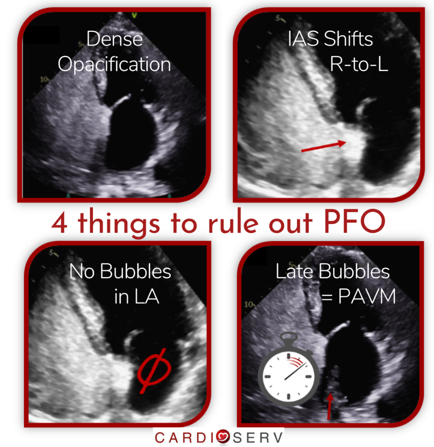
A Patent Foramen Ovale (PFO) It is a slit-like defect resulting from an incomplete fusion of the foramen ovale within the atrial septum. 20-25% of the population have a PFO and echocardiography is often used to diagnosis it. This week we will review the 4 things needed to rule out a PFO during an...
 Cardioserv Blog